


Validation of the non-sentinel lymph node metastasis prediction models in Chinese breast cancer population and the construction of a new model
-
摘要:
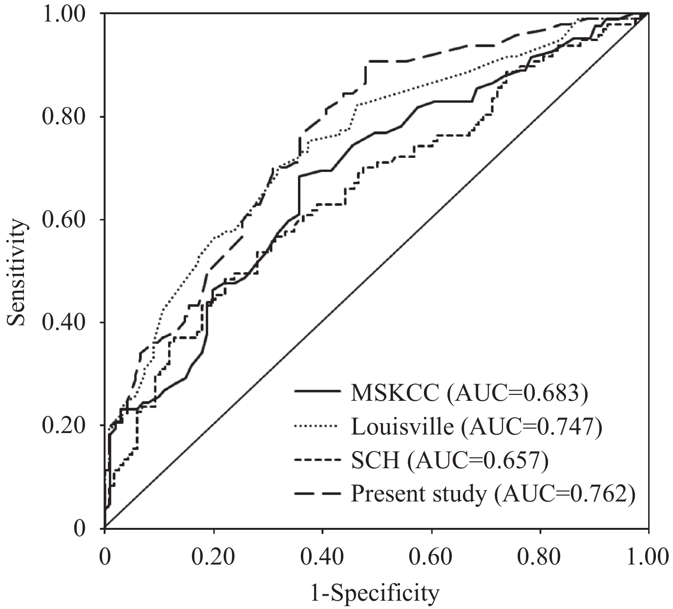
目的 评估纪念斯隆-凯特琳癌症中心(MSKCC)、Louisville和SCH等3种乳腺癌非前哨淋巴结(non-SLN)转移风险预测模型在中国人群中的临床应用价值,并构建新的non-SLN转移风险预测模型。 方法 本研究共纳入220例在我院进行SLN活检阳性和腋窝淋巴结清扫的乳腺癌患者。对患者的临床病理资料进行单因素分析和多因素logistic回归分析,根据筛选出的non-SLN的独立预测因素建立1个新的non-SLN转移风险预测模型。使用所纳入的乳腺癌患者临床病理资料对MSKCC、Louisville和SCH以及新建立的模型进行比较,绘制受试者工作特征曲线,计算各模型的曲线下面积及假阴性率,以评估各模型的区分能力和临床应用价值。 结果 所纳入的220例中国乳腺癌患者中,有97例(44.1%)发现腋窝淋巴结转移阳性。经logistic回归分析,发现原发肿瘤大小、脉管癌栓有无、阳性SLN的数量以及阳性SLN /总SLN的比值是non-SLN转移的独立预测因素,并据此建立了1个新的non-SLN转移风险预测模型。在预测non-SLN转移风险时,MSKCC、Louisville、SCH以及新的模型的曲线下面积分别为0.683、0.747、0. 657和0.762。各模型的假阴性率调整到接近10%时,MSKCC、Louisville、SCH和新建模型分别将17.27%、20.00%、15.00% 和33.18%的病人归类到低风险组中。 结论 与MSKCC、Louisville模型和SCH模型比较,本研究建立的新模型在对乳腺癌non-SLN转移风险的预测方面可能具有更高的临床价值,但在应用此模型前仍需更大样本的资料进行验证和修正。对于SLN活检阳性的乳腺癌患者,在应用非前哨淋巴结转移风险预测模型辅助是否进行腋窝淋巴结清扫的临床决策时需谨慎。 Abstract:Objective To assess the clinical values of 3 non-sentinel lymph node (SLN) metastasis prediction models, including MSKCC, Louisville and SCH, in Chinese population, and to construct a new model for predicting the risk of non-sentinel lymph node metastasis. Methods A total of 220 breast cancer patients who underwent SLN biopsy and axillary lymph node dissection (ALND) in our hospital were included in this study. After performing a univariate analysis and multivariate logistic regression analysis on the clinicopathological data of the patients, a new non-SLN metastasis risk prediction model was established based on the independent predictors we identified. MSKCC, Louisville, SCH and the newly established model were compared using the clinicopathological data of our patients, after which the receiver operating characteristic curve (ROC) was plotted and the area under the curve (AUC) and false negative rates (FNR) of each model was calculated to evaluate the discrimination capability and clinical value of above models. Results Of the 220 breast cancer patients included, 97 patients (44.1%) were found to have positive non-SLNs. Logistic regression analysis indicated that only the size of the primary tumor size, vessel cancerous emboli, number of positive SLN and proportion of positive SLN/total SLN were independent predictors of non-SLN metastasis, based on which a new prediction model was constructed. The AUC values of the MSKCC, Louisville, SCH and the new model were 0.683, 0.747, 0.657 and 0.762 respectively. When FNR was adjusted close to 10%, the percentage of patients who were classified into the low-risk group by MSKCC, Louisville, SCH and the new model were 17.27%, 20.00%, 15.00% and 33.18%. Conclusion The new model established in this study may be more clinically valuable in predicting the risk of non-SLN metastasis than MSKCC, Louisville and SCH, but before its application a larger sample is needed for model validation and adjustment. Doctors should be cautious when using non-SLN metastasis predicting models for decision making as whether patients with positive SLN biopsy should complete ALND. -
Key words:
- breast cancer /
- prediction models /
- sentinel lymph node /
- non-sentinel lymph node metastasis /
- nomogram
-
表 1 乳腺癌患者临床病理特征的单变量分析
特征 非前哨淋巴结转移[n (%)] P 阴性 阳性 病例 123(55.9%) 97(44.1%) 年龄(岁) 0.655 ≤50 75 (61.0%) 62 (63.9%) >50 48 (39.0%) 35 (36.1%) 钼靶 0.074 阴性 99(80.5%) 68(70.1%) 阳性 24(19.5%) 29(29.9%) 原发灶大小 0.003 T1 68 (55.3%) 38 (39.2%) T2 53 (43.1%) 47 (48.5%) T3 2 (1.6%) 12 (12.4%) 多灶性 0.309 是 6(4.9%) 8 (8.2%) 否 117 (95.1%) 89 (91.8%) 原发肿瘤位置 0.928 外上象限 39 (31.37%) 35 (36.1%) 外下象限 14 (11.4%) 35 (36.1%) 内上象限 16 (13.0%) 29 (29.9%) 内下象限 9 (7.3%) 15(15.5%) 中央区或跨象限 45 (36.6%) 12(12.4%) 病理类型 0.773 浸润性导管癌 112 (91.1%) 87 (89.7%) 浸润性小叶癌 3 (2.4%) 5 (5.2%) 其他 8 (6.5%) 5 (5.2%) 病理学分级 0.016 I级 7 (5.7%) 2 (2.1%) II级 62 (50.4%) 37 (38.1%) III级 42 (34.1%) 46 (47.4%) 未知 12(9.8%) 12 (12.4%) 淋巴血管侵犯 <0.001 有 33(26.8%) 49 (50.5%) 无 90 (73.2%) 48 (49.5%) ER 0.762 阳性 91 (74.0%) 70 (72.2%) 阴性 32 (26.0%) 27(27.8%) PR 0.815 阳性 88 (71.5%) 68 (70.1%) 阴性 35(28.5%) 29 (29.9%) Her-2 0.140 阳性 29 (23.6%) 29 (29.9%)  下载: 导出CSV
表 2 乳腺癌患者non-SLN转移风险因素的Logistic回归分析结果
变量 系数 标准误 Wals P OR 95% 置信区间 下界值 上界值 原发灶大小 (vsT1) 6.076 0.048 T2 0.273 0.314 0.756 0.385 1.314 0.710 2.429 T3 2.073 0.854 5.891 0.015 7.945 1.490 42.363 淋巴血管侵犯 0.723 0.321 5.085 0.024 2.061 1.099 3.866 转移阳性的前哨淋巴结个数 0.377 0.149 6.416 0.011 1.458 1.089 1.952 转移阳性的前哨淋巴结个数/前哨淋巴结总个数 1.780 0.511 12.139 0.000 5.930 2.179 16.142 常量 –2.427 0.422 33.093 0.000 0.088
下载: 导出CSV
表 3 乳腺癌患者non-SLN转移风险预测模型的AUC值和假阴性率比较
模型 验证 调整 AUC(95%置信区间) FNR值* 低于原阈值且非前
哨淋巴结转移阳性
的患者个数低于原阈值
的患者总数调整后FNR值** 低于调整后阈值且
非前哨淋巴结转移
阳性的患者个数低于调整后阈值
的患者总数MSKCC 2.44% 2 11(6.01%) 9.27% 9 38(17.27%) 0.683(0.606-0.761) Louisville 0.00% 0 0(0%) 9.27% 9 44(20.00%) 0.747(0.682-0.812) SCH 0.00% 0 0(0%) 9.27% 9 33(15.00%) 0.657(0.583-0.730) Present study - - - 9.27% 9 73(33.18%) 0.762(0.700-0.825) *FNR值=低于原阈值且非前哨淋巴结转移阳性的患者个数/低于原阈值的患者总数; **调整后FNR值=低于调整后阈值且非前哨淋巴结转移阳性的患者个数/低于调整后阈值的患者总数.
下载: 导出CSV
-
[1] Choi HY, Park M, Seo M, et al. Preoperative axillary lymph node evaluation in breast cancer current issues and literature review[J]. Ultrasound Q, 2017, 33(1): 6-14. doi: 10.1097/RUQ.0000000000000277 [2] Krag DN, Weaver DL, Alex JC, et al. Surgical resection and radiolocalization of the sentinel lymph node in breast cancer using a gamma probe[J]. Surg Oncol, 1993, 2(6): 335-9. doi: 10.1016/0960-7404(93)90064-6 [3] 杨翼鹏, 嵇健, 刘民锋, 等. 腋窝治疗对于早期乳腺癌患者是否有必要[J]. 南方医科大学学报, 2014, 34(7): 1065-7. [4] Veronesi U, Paganelli G, Galimberti V, et al. Sentinel-node biopsy to avoid axillary dissection in breast cancer with clinically negative lymph-nodes[J]. Lancet, 1997, 349(969): 1864-7. [5] Chen SL, Iddings DM, Scheri RP, et al. Lymphatic mapping and sentinel node analysis: current concepts and applications[J]. CA Cancer J Clin, 2006, 56(5): 292-309. doi: 10.3322/canjclin.56.5.292 [6] Poortmans PM, Struikmans H, Bartelink H. Regional nodal irradiation in Early-Stage breast cancer[J]. N Engl J Med, 2015, 373(19): 1879-80. [7] Speers C, Pierce LJ. Postoperative radiotherapy after Breast-Conserving surgery for Early-Stage breast cancer a review[J]. JAMA Oncol, 2016, 2(8): 1075-82. doi: 10.1001/jamaoncol.2015.5805 [8] Garcia-Tejedor A, Falo C, Quetglas CA, et al. Feasibility, accuracy and prognosis of sentinel lymph node biopsy before neoadjuvant therapy in breast cancer. A prospective study[J]. Int J Surg, 2017, 39(11): 141-7. https://www.sciencedirect.com/science/article/pii/S1743919117301115 [9] Manca G, Rubello D, Tardelli E, et al. Sentinel lymph node biopsy in breast cancer:indications,contraindications,and controversies[J]. Clin Nucl Med, 2016, 41(2): 126-33. doi: 10.1097/RLU.0000000000000985 [10] Chen JY, Chen JJ, Yang BL, et al. Predicting sentinel lymph node metastasis in a Chinese breast cancer population: assessment of an existing nomogram and a new predictive nomogram[J]. Breast Cancer Res Treat, 2012, 135(3): 839-48. doi: 10.1007/s10549-012-2219-x [11] Chagpar AB, Scoggins CR, Martin IR, et al. Prediction of sentinel lymph node-only disease in women with invasive breast cancer[J]. Am J Surg, 2006, 192(6): 882-6. doi: 10.1016/j.amjsurg.2006.08.063 [12] Qiu PF, Liu JJ, Wang YS, et al. Risk factors for sentinel lymph node metastasis and validation study of the MSKCC nomogram in breast cancer patients[J]. Jpn J Clin Oncol, 2012, 42(11): 1002-7. doi: 10.1093/jjco/hys150 [13] Van Zee KJ, Manasseh DM, Bevilacqua JL, et al. A nomogram for predicting the likelihood of additional nodal metastases in breast cancer patients with a positive sentinel node biopsy[J]. Ann Surg Oncol, 2003, 10(10): 1140-51. doi: 10.1245/ASO.2003.03.015 [14] Weaver DL, Ashikaga T, Krag DN, et al. Effect of occult metastases on survival in node-negative breast cancer[J]. N Engl J Med, 2011, 364(5): 412-21. doi: 10.1056/NEJMoa1008108 [15] Barranger E, Coutant C, Flahault A, et al. An axilla scoring system to predict non-sentinel lymph node status in breast cancer patients with sentinel lymph node involvement[J]. Breast Cancer Res Treat, 2005, 91(2): 113-9. doi: 10.1007/s10549-004-5781-z [16] 吴佩琪, 刘春玲, 刘再毅, 等. 钼靶、CT与DCE-MRI评价乳腺癌淋巴结转移的价值[J]. 南方医科大学学报, 2016, 36(4): 493-9. [17] Singletary SE, Greene FL, Sobin LH. Classification of isolated tumor cells:clarification of the 6th edition of the American Joint Committee on cancer staging manual[J]. Cancer, 2003, 98(12): 2740-1. doi: 10.1002/(ISSN)1097-0142 [18] Kim WG, Lee JS. Axillary skip metastases and the False-Negative rate of sentinel lymph node biopsy in patients with breast cancer are related to negative ALDH-1 expression and Ki-67 expression[J]. Int J Surg Pathol, 2017, 25(5): 397-405. doi: 10.1177/1066896917690024 [19] Kohrt HE, Olshen RA, Bermas HR, et al. New models and online calculator for predicting non-sentinel lymph node status in sentinel lymph node positive breast cancer patients[J]. BMC Cancer, 2008, 8(12): 66-75. doi: 10.1186/1471-2407-8-66 [20] Cho J, Han W, Lee JW, et al. A scoring system to predict nonsentinel lymph node status in breast cancer patients with metastatic sentinel lymph nodes: A comparison with other scoring systems[J]. Ann Surg Oncol, 2008, 15(8): 2278-86. doi: 10.1245/s10434-008-9993-z [21] Perhavec A, Perme MP, Hocevar M, et al. Ljubljana nomograms for predicting the likelihood of non-sentinel lymph node metastases in breast cancer patients with a positive sentinel lymph node[J]. Breast Cancer Res Treat, 2010, 119(2): 357-66. doi: 10.1007/s10549-009-0561-4 [22] Gur AS, Unal B, Ozbek U, et al. Validation of breast cancer nomograms for predicting the non-sentinel lymph node metastases after a positive sentinel lymph node biopsy in a multi-center study[J]. Eur J Surg Oncol, 2010, 36(1): 30-5. doi: 10.1016/j.ejso.2009.05.007 [23] Huang J, Chen X, Fei X, et al. Risk factors of non-sentinel lymph node metastasis and performance of MSKCC nomogramin breast cancer patients with metastatic sentinel lymph node[J]. Chin J Surg, 2015, 53(12): 941-6. [24] 陈嘉莹, 陈嘉健, 杨犇龙, 等. MSKCC乳腺癌前哨淋巴结转移预测模型的验证性研究[J]. 中国实用外科杂志, 2011, 28(01): 89-93. http://www.cqvip.com/QK/90965A/201101/36429809.html [25] 黄佳慧, 陈小松, 费晓春, 等. 乳腺癌非前哨淋巴结转移危险因素及MSKCC模型预测效能研究[J]. 中华外科杂志, 2015, 53(12): 941-6. doi: 10.3760/cma.j.issn.0529-5815.2015.12.011 [26] 曹迎明, 刘淼, 周波, 等. MSKCC和SOC模型预测中国乳腺癌患者非前哨淋巴结转移的验证比较研究[J]. 中国肿瘤临床, 2014,33(08): 508-12. doi: 10.3969/j.issn.1000-8179.21031009 [27] 邱鹏飞, 刘雁冰, 王永胜, 等. 乳腺癌前哨淋巴结转移相关因素与MSKCC列线图的验证性研究[J]. 中华内分泌外科杂志, 2012, 06(5): 307-12. http://d.wanfangdata.com.cn/Periodical_nfmwk201205006.aspx [28] 罗铭. 乳腺癌非前哨淋巴结转移风险预测模型的验证及新模型的建立[D]. 南宁: 广西医科大学, 2014. [29] 陈嘉莹. 早期乳腺癌前哨淋巴结及非前哨淋巴结转移预测模型的建立和验证[D]. 上海: 复旦大学, 2012. -

 点击查看大图
图(1) / 表(3)
计量
- 文章访问数: 1587
- HTML全文浏览量: 565
- PDF下载量: 2
- 被引次数: 0