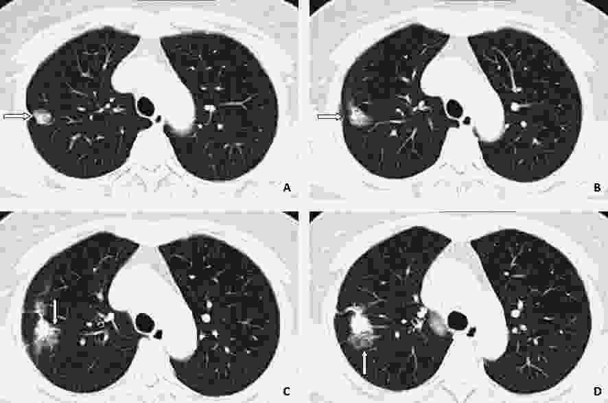
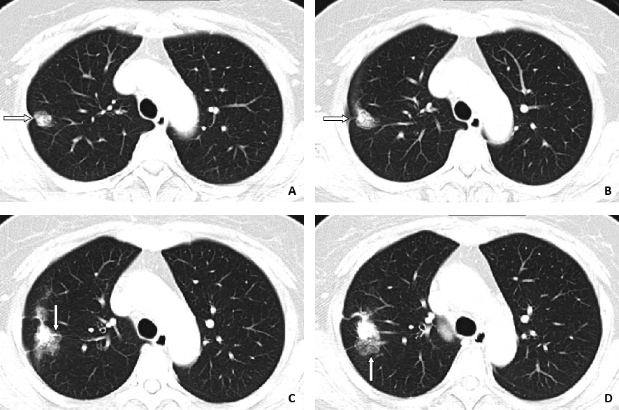
Value of various CT findings in the differential diagnosis of peripheral lung cancer and tuberculosis
-
摘要:
目的 探讨周围型肺癌与肺结核的有价值CT征象。 方法 回顾性分析经病理证实的58例肺癌及32例肺结核患者的多样化CT征象,肺癌为病例组,肺结核为对照组,提取有价值征象进行比较并归纳总结。 结果 肺癌组有腺癌46例、鳞癌8例、小细胞癌4例,主要征象分别是:分叶49(84.5%)例,毛刺39(67.2%)例,胸膜凹陷20(34.4%)例,晕征13(22.4%)例;肺结核组主要征象分别是:胸膜反应26(81.25%)例,卫星灶22(68.75%)例,钙化22(68.75%)例,毛刺26(81.25%)例。肺癌与肺结核组CT征象比较中,分叶、晕征、胸膜凹陷、钙化、卫星灶、胸膜反应等征象的分布差异均有统计学意义(P<0.05),而两者CT征象诊断准确度的比较中,肺癌分叶的诊断指数>150%,肺结核钙化、卫星灶、胸膜反应的诊断指数均>150%。 结论 周围型肺癌与肺结核的鉴别诊断中,分叶、胸膜凹陷、晕征、钙化、卫星灶、胸膜反应等CT征象对于肺癌与肺结核的诊断有一定临床价值。 Abstract:Objective To explore the value of various CT findings of peripheral lung cancer and tuberculosis. Methods Diverse CT findings of 58 cases of lung cancer and 32 cases of pulmonary tuberculosis which was confirmed by pathology were retrospective analyzed. Patient with lung cancer was case group, and patient with tuberculosis was control group. Valuable signs were extracted and compared between 2 groups. Results Lung cancer group had 46 cases of adenocarcinoma, 8 cases of squamous cell carcinoma and 4 cases of small cell carcinoma.The main signs were: 49 cases(84.5%)with lobulate, 39(67.2%) with burr, 20 cases(34.4%) with pleural indentation, 13(22.4%) with halo sign .In main signs of tuberculosis,pleural reaction was 26 (81.25%), the satellite focal point 22 (68.75%), calcification 22 (68.75%), burr 26 (81.25%).Difference of lobulate, halo, pleural depression, calcification, satellite focal point and pleural reaction between lung cancer group and tuberculosis group were significant (P<0.05). The diagnostic index of lobule of lung cancer was >150%, the diagnostic indexes of tuberculosis calcification、satellite focal area and pleural membrane reaction were all > 150%, which had a diagnostic value. Conclusion In the differential diagnosis of patients with peripheral lung cancer and tuberculosis, the CT signs such as lobulate, pleural depression, halo, calcification, satellite focal point and pleural reaction have a certain clinical value for the diagnosis of ung cancer and tuberculosis. -
Key words:
- diversity signs of CT findings /
- peripheral lung cancer /
- CT scan /
- diagnostic value
-
表 1 肺癌与肺结核多样化CT征象检验结果(n)
征象 肺癌 肺结核 χ2 P 分叶 有 49 4 44.137 0.000 无 9 28 毛刺 有 39 26 2.017 0.220 无 19 6 胸膜凹陷 有 20 3 6.833 0.011 无 38 29 晕征 有 13 0 8.383 0.003 无 45 32 远侧阻塞性肺炎 有 14 3 2.934 0.100 无 44 29 蜘蛛腿征 有 13 3 2.399 0.156 无 45 29 钙化 有 1 22 48.697 0.000 无 57 10 卫星灶 有 7 22 30.337 0.000 无 51 10 胸膜反应 有 4 26 51.304 0.000 无 54 6 近端支气管梗阻扩张 有 9 3 0.673 0.527 无 49 29 表 2 肺癌和肺结核组多样化CT征象诊断准确度评价
征象 灵敏度(%) 特异度(%) 诊断指数(%) Youden指数(%) 肺癌 肺结核 肺癌 肺结核 肺癌 肺结核 肺癌 肺结核 分叶 84.48 12.51 87.5 15.51 171.98 28 71.98 -72 毛刺 67.24 81.25 18.7 32.75 85.99 114 -14.06 14 胸膜凹陷 34.48 9.37 90.62 61.51 125.1 74.88 25.1 -25.12 晕征 22.41 0 100 77.59 122.41 77.59 22.41 -22.41 远侧阻塞性肺炎 24.13 9.37 90.62 75.86 114.75 85.23 14.75 -14.77 钙化 1.72 68.75 31.25 98.27 32.97 167.02 -67.03 67.02 卫星灶 12.06 68.75 31.25 87.93 43.31 156.68 -56.69 56.68 胸膜反应 6.89 81.25 18.75 93.1 25.64 174.35 -74.36 74.35 -
[1] 白人驹, 张雪林. 医学影像诊断学[M]. 北京: 人民卫生出版社, 2014. [2] Lemjabbar-Alaoui H, Hassan O, Yang YW, et al. Lung cancer: biology and treatment options[J]. Biochim Biophys Acta, 2015, 1856(2): 189-210. https://www.sciencedirect.com/science/article/pii/S0304419X15000669 [3] 马跃虎, 李永霞, 杨帆. 周围型小肺癌的CT表现与病理对照分析[J]. 中国现代医学杂志, 2013, 23(31): 100-3. doi: 10.3969/j.issn.1005-8982.2013.31.026 [4] Ebara K, Takashima S, Jiang BH, et al. Pleural invasion by peripheral lung cancer: prediction with Three-Dimensional CT[J]. Acad Radiol, 2015, 22(3): 310-9. doi: 10.1016/j.acra.2014.10.002 [5] 孙泽源, 何志颖, 梁培生. 90例周围型肺癌的影像学表现[J]. 广东医学, 2011, 32(6): 774-5. http://d.wanfangdata.com.cn/Periodical_gdyx201106038.aspx [6] 张定均. 周围型肺癌100例CT图像特征分析[J]. 现代中西医结合杂志, 2013, 22(6): 653-4. http://med.wanfangdata.com.cn/Paper/Detail/PeriodicalPaper_shandyy201203023 [7] 张露钢, 郑红斌. 球型肺结核与周围型肺癌的CT征象对比研究[J]. 中国现代医生, 2013, 51(11): 92-4. [8] 郑勇, 孔江明, 高文军. 螺旋CT扫描对周围型肺癌的诊断价值[J]. 中国基层医药, 2011, 18(20): 2798-9. doi: 10.3760/cma.j.issn.1008-6706.2011.20.034 [9] 杨帆, 马跃虎, 陈跃芳, 等. 早期周围型肺癌96例CT影像学特点分析[J]. 山东医药, 2012, 52(3): 64-5. https://www.wenkuxiazai.com/doc/0b346b106bd97f192279e94c.html [10] 田晓敏, 卢智. 球形肺结核与周围型肺癌的CT诊断与临床分析[J]. 医学影像学杂志, 2014, 24(8): 1414-6. [11] 张超, 张俊祥, 刘德武, 等. 周围型肺癌螺旋CT征象与p53、VEGF表达的关系[J]. 山东医药, 2013, 53(12): 47-9. doi: 10.3969/j.issn.1002-266X.2013.12.019 [12] 李辉, 阚晓婧, 宁培刚, 等. HRCT常见恶性征象对孤立性肺结节的定性诊断[J]. 放射学实践, 2014, 29(12): 1405-8. http://med.wanfangdata.com.cn/Paper/Detail/PeriodicalPaper_fsxsj201412015 [13] 何亚奇, 唐秉航, 林乐, 等. 肺腺癌浸润前病变的CT诊断[J]. 放射学实践, 2017, 32(8): 835-8. http://www.cqvip.com/QK/98509X/201404/662244052.html [14] 韩瑜, 王振光, 刘思敏, 等. 胸膜凹陷MSCT和18F-FDGPET/CT特征评价周围型肺癌胸膜侵犯[J]. 中国医学影像技术, 2014, 29(12): 1835-8. http://d.wanfangdata.com.cn/Thesis/Y2587218 [15] 曹捍波, 王梅, 王和平. MSCT对孤立性肺结节(≤2 cm)胸膜凹陷征的诊断及鉴别诊断价值[J]. 医学影像学杂志, 2017, 27(8): 1471-4. http://www.cqvip.com/QK/96184X/201306/47998264.html [16] 韩瑜, 王振光, 刘思敏, 等. 胸膜凹陷征MSCT和18F-FDG PET/CT特征评价周围型肺癌胸膜侵犯[J]. 中国医学影像技术, 2014, 30(12): 1835-8. http://journal.9med.net/html/qikan/yykxzh/hbyy/200993118/lz/20100329094828149_520134.html [17] 肖湘生, 吴华伟, 李惠民, 等. 周围型肺癌胸膜凹陷的CT和MRI表现与病理对照[J]. 临床放射性杂志, 2002, 21(5): 344-7. [18] Rubin P, Hansen JT. Tnm staging atlaswith oncoanatomy[J]. Lippincott Williams Wilkins, 2011, 38(4): 185-8. http://www.doc88.com/p-7714993043092.html [19] 侯唯姝, 殷焱, 程杰军, 等. 能谱CT成像在鉴别周围型肺癌和肺炎性肿块中的价值[J]. 中华放射学杂志, 2014, 48(10): 832-5. doi: 10.3760/cma.j.issn.1005-1201.2014.10.010 [20] 周坦峰, 吴伟. 周围型肺癌与结核瘤影像诊断及鉴别诊断的临床研究[J]. 临床肺科杂志, 2016, 21(5): 958-60. http://www.cqvip.com/QK/83745X/201605/668490466.html [21] 卫惠芳, 陈菊花. 酷似肺癌的肺结核46例临床分析[J]. 中国防痨杂志, 2011, 33(5): 283-4. [22] 范丽, 望云, 管宇, 等. 临床I期周围型肺癌的MDCT及误诊原因分析[J]. 临床放射学杂志, 2016, 35(3): 354-9. http://www.cnki.com.cn/Article/CJFDTOTAL-LCFS201603012.htm -


 下载:
下载:


