


Diffusion weighted magnetic resonance imaging combined with dynamic enhancement technique in identification of benign and malignant breast lesions
-
摘要:
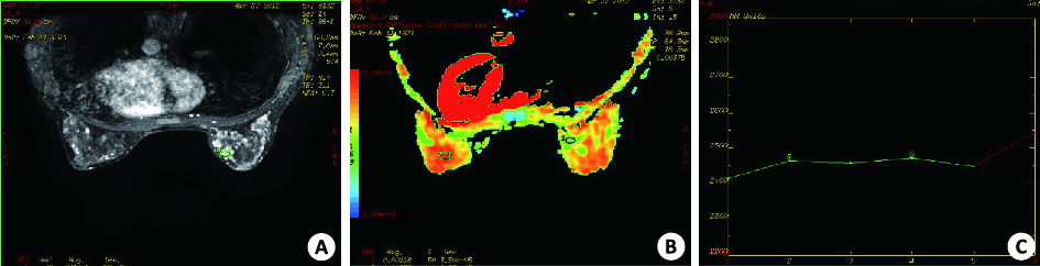
目的 评估磁共振弥散加权成像(MR-DWI)、动态增强成像(DCE-MRI)联合鉴别乳腺良恶性肿块的价值。 方法 回顾性分析2012年1月~2015年12月期间经病理证实的女性乳腺肿块患者70名。所有患者均行MR-DWI及DCE-MRI,诊断结果与病理进行对照。 结果 70例患者共77例病灶。25例恶性肿块,动态增强Ⅰ型曲线3例(12%),Ⅱ型曲线7例(28%),Ⅲ型曲线15例(60%);52例良性肿块,动态增强Ⅰ型曲线35例(67%),Ⅱ型曲线11例(21%),Ⅲ型曲线6例(12%)。良恶性肿块的Ⅰ型及Ⅲ型曲线的差异均有统计学意义(P<0.05)。良恶性肿块的表观弥散系数(ADC)差异亦有统计学意义(P=0.000)。ROC曲线分析,动态增强曲线的敏感性和特异性分别为85%和60%,曲线下面积(AUC)为0.723;ADC值诊断乳腺良恶性肿块的敏感性和特异性分别为88%和78%,AUC为0.830;联合ADC值、动态增强曲线敏感性和特异性分别为90%和98%,AUC为0.943。 结论 DWI的ADC值较DCE-MRI具有更高的敏感性;联合应用DWI、DCE-MR可提高鉴别乳腺肿块良恶性的敏感性和特异性。 Abstract:Objective To assess the value of diffusion weighted magnetic resonance imaging combined with dynamic enhancement technique in identification of benign and malignant breast lesions. Methods Seventy patients with breast lumps confirmed by pathology were retrospectively analyzed.The patients were underwent with DW imaging examination and dynamic contrast-enhanced imaging. The diagnosis result were compared with pathologic results. Results The 70 patients had 77 lesions. DCE-MRI had 3(12%) typeⅠcurving patterns, 7(28%) typeⅡ curving patterns and 15(60.0%) type Ⅲ curving patterns in the malignant group.In benign group, there were 35(67%) typeⅠcurving patterns, 11(21%) type Ⅱ curving patterns and 6(12%) type Ⅲ curving patterns. Curving pattern of TypeⅠand type Ⅲ were significantly different (P<0.05). The difference of ADC values between benign and malignant tumor was significant (P=0.000). The curve of ROC analysis showed DCE-MRI with 85.0% sensitivity, 60% specificity and 0.723 AUC. The sensitivity, specificity, AUC of ADC were 88%, 78% and 0.83, respectively. The combination of two technique had sensitivity of 90%, specificity of 98% and AUC of 0.943. Conclusion Compared to DCE-MRI, ADC have a high accuracy in diagnosis of benign and malignant breast tumor. The application of DWI and DEC-MRI can improve the accuracy of diagnosis of breast tumor. -
表 1 良恶性肿块动态增强TIC类型(n,%)
病理类型 n Ⅰ型(流入型) Ⅱ型(平台型) Ⅲ型(流出型) 恶性肿块 25 3(12) 7(28) 15(60) 良性肿块 52 35(67) 11(21) 6(12) P <0.05 >0.05 <0.05 χ2 21.1 0.19 18.9  下载: 导出CSV
表 2 ADC、DEC-MRI及两者联合诊断的分析指标
检查方法 曲线下面积 敏感性(%) 特异性(%) ADC 0.830* 88 78 DEC-MRI 0.723* 85 60 两者联合 0.943 90 98 *P<0.05vs两者联合; ADC: 表观扩散系数; DEC-MRI: 动态增强成像.
下载: 导出CSV
-
[1] Chen X, Li WL, Zhang YL, et al. Meta-analysis of quantitative diffusion-weighted Mr imaging in the differential diagnosis of breast lesions[J]. BMC Cancer, 2010, 29(10): 693-6. [2] Fornasa F, Pinali L, Gasparini A, et al. Diffusion-weighted magnetic resonance imaging in focal breast lesions: analysis of 78 casts with pathological correlation[J]. Radiol Med, 2011, 116(2): 264-75. doi: 10.1007/s11547-010-0602-4 [3] 徐茂林, 谢东, 康巍, 等. DCE-MRI结合DWI对乳腺NMLE良恶性病变的鉴别诊断价值[J]. 中国CT和MRI杂志, 2015, 24(8): 43-6. http://d.wanfangdata.com.cn/Periodical/zgcthmrizz201508014 [4] 靳雅楠, 张焱, 程敬亮, 等. DCE-MRI及DWI在鉴别乳腺良, 恶性病变中的价值[J]. 郑州大学学报: 医学版, 2016, 51(4): 530-3. http://d.wanfangdata.com.cn/Periodical/henanykdx201604025 [5] 张小安, 刘真真, 赵鑫, 等. 动态增强MRI结合DWI对乳腺病变性质的诊断价值[J]. 实用放射学杂志, 2013, 29(4): 561-4. http://d.wanfangdata.com.cn/Periodical/syfsxzz201304014 [6] 阳君, 苏丹柯, 赵欣, 等. 动态增强磁共振联合扩散加权成像技术对乳腺环形强化病变的诊断应用价值研究[J]. 临床放射学杂志,2016, 35(10): 1490-4. http://d.wanfangdata.com.cn/Periodical/lcfsxzz201610011 [7] 张丽, 韩立新, 曹惠霞, 等. 3.0T磁共振扩散加权成像和VIBRANT动态增强在鉴别乳腺腺病与乳腺癌中的价值[J].临床放射学杂志, 2017, 36(3): 342-6. [8] 罗禹, 王培军, 周永明, 等. 磁共振动态增强灌注成像参数图在乳腺良恶性病变诊断中的价值[J]. 医学影像学杂志, 2016, 26(4): 649-54. http://d.wanfangdata.com.cn/Periodical/yxyxxzz201604027 [9] 姚薇薇, 夏军, 杜恒峰, 等. 基于动态增强磁共振的T1WI定量灌注参数伪彩图在导管源性乳腺癌中的诊断价值[J]. CT理论与应用研究, 2014, 23(1): 131-8. http://d.wanfangdata.com.cn/Periodical/ctta201401015 [10] Tozaki M, Fukuda K. High-spatial-resolution MRI of non-masslike breast lesions: interpretation model based on BI-RADS MRI descriptors[J]. AJR Am J Roentgenol, 2006, 187(2): 330-7. doi: 10.2214/AJR.05.0998 [11] Inoue K, Kozawa E, Mizukoshi W, et al. Usefulness of diffusion-weighted imaging of breast tumors: quantitative and visual assessment[J]. Jpn J Radiol, 2011, 29(6): 429-36. doi: 10.1007/s11604-011-0575-9 [12] 彭艳霞, 蔡宏民, 崔春艳, 等. DWI及动态增强MRI鉴别乳腺病变的对比研究[J]. 中国CT和MRI杂志, 2014, 11(1): 1-4. http://d.wanfangdata.com.cn/Periodical/zgcthmrizz201401001 [13] 王宇翔, 刘金芝, 刘欢, 等. 磁共振扩散加权成像联合动态增强在乳腺良恶性病变鉴别诊断中的价值[J]. 实用临床医药杂志, 2017, 21(1): 136-8. http://d.wanfangdata.com.cn/Periodical/jslcyxzz201701046 [14] Chen X, He XJ, Jin R, et al. Conspicuity of breast lesions at different b values on diffusion-weighted imaging[J]. BMC Cancer, 2012, 2(12): 334-7. [15] Knopp MV, Bourne MW, Sardanelli F, et al. Gadobenate dimeglumine-enhanced MRI of the breast: analysis of dose response and comparison with gadopentetate dimeglumine[J]. AJR Am J Roentgenol, 2003, 181(3): 663-76. doi: 10.2214/ajr.181.3.1810663 [16] Szabo BK, Aspelin P, Wiberg MK, et al. Dynamic Mr imaging of the breast: analysis of kinetic and morphologic diagnostic criteria[J]. Acad Radiol, 2003, 14(9): 379-86. [17] 朱融, 董光, 耿海, 等. 动态增强磁共振扫描及弥散加权成像对无占位效应乳腺病灶的诊断价值[J]. 医学影像学杂志, 2015, 14(6): 1083-7. http://d.wanfangdata.com.cn/Periodical/yxyxxzz201506045 [18] 程赛楠, 刘维娜. 磁共振扩散加权成像在乳腺肿瘤中的临床应用[J]. 医学影像学杂志, 2014, 24(1): 129-32. http://d.wanfangdata.com.cn/Periodical/yxyxxzz201401040 [19] 吴春莲, 武彤彤, 蔡方, 等. 乳腺导管内增生性病变中Survivin的表达及意义[J]. 分子影像学杂志, 2014, 37(2): 100-2. http://d.wanfangdata.com.cn/Periodical/fzyxxzz201402011 [20] 周礼金, 李晓杰, 纪婷. 磁共振动态增强VIEWS及弥散加权成像DWI在乳腺癌保乳术前评估的价值[J]. 中国CT和MRI杂志, 2015, 21(5): 74-6, 110. http://d.wanfangdata.com.cn/Periodical/zgcthmrizz201505023 [21] Imamura T, Isomoto I, Sueyoshi E, et al. Diagnostic performance of ADC for Non-mass-like breast lesions on Mr imaging[J]. Magn Reson Med Sci, 2010, 9(4): 217-25. doi: 10.2463/mrms.9.217 -

 点击查看大图
图(1) / 表(2)
计量
- 文章访问数: 656
- HTML全文浏览量: 270
- PDF下载量: 9
- 被引次数: 0