


Progress of artificial intelligence in the field of pathological diagnosis
-
摘要: 基于近几年机器视觉的发展,深度学习的人工智能方法应用于组织病理极大程度上促进了病理学家解决临床上的诊断问题,用该种方法解决病理学问题可被称为计算机病理学。人工智能可以做到帮助病理学家初筛大部分良性数据、辅助诊断、疗效预测、识别生物标志物等,甚至可以做到对药效治疗监测以及识别药物发现未知的信号。基于深度学习在病理领域的深入研究,让计算机自动处理病理数据成为可能。人工智能诊断决策建立在大数据之上,很多有可能做到对每个病人的个性化管理,对于大多普遍性的疾病诊断有着更加快速准确的优势。但数字病理学的发展仍受到一些问题的限制,以至于现阶段没有广泛应用于数字病理诊断平台。本文总结了近几年人工智能在病理诊断领域的最新进展,并讨论这种技术的可行性,补充说明在数字病理学中遇到的困难和挑战,并提出在该领域实用性上的展望。Abstract: Based on the development of machine vision in recent years, the application of deep learning artificial intelligence methods in histopathology has greatly promoted pathologists to solve clinical diagnostic problems. This method can be called computer pathology. Artificial intelligence can help pathologists sift through most benign data, aid in diagnosis, predict efficacy, identify biomarkers, and even monitor therapeutic efficacy and identify unknown signals for drugs. Based on the in-depth study of deep learning in the field of pathology, it is possible for the computer to process pathological data automatically. Artificial intelligence diagnostic decisions are based on big data, and it is possible to personalize the management of each patient. It has the advantage of more rapid and accurate diagnosis for most common diseases. However, it is worth considering that the development of digital pathology is still limited by some problems, so that it is not widely used in digital pathology diagnostic platform at the present stage. We summarized the recent progress of artificial intelligence in the field of pathological diagnosis in this paper. We discussed the feasibility of this technology, added the difficulties and challenges encountered in digital pathology, and put forward the prospect of its practicality in this field.
-
Key words:
- digital pathological /
- computer-aided diagnosis /
- deep learning /
- whole slide image
-

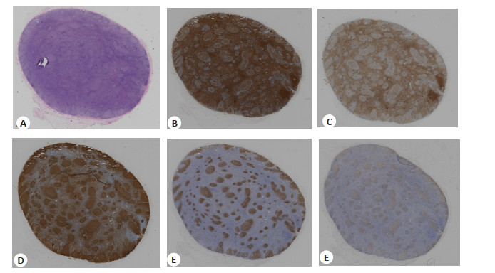
图 1 同一淋巴结病理切片
A: H & E; B: 免疫组化CD3; C: 免疫组化CD5; D: 免疫组化CD20; E: 免疫组化CD21; F: 免疫组化CD23.
Figure 1. Pathological sections of the same lymph node.

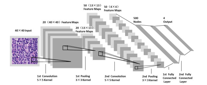
图 2 用于检测图像中视觉类别的卷积神经网络的处理流程
Figure 2. Processing pipeline of convolution alneural network for the detection of visualcategoriesin images.
-
[1] Araújo ALD, Arboleda LPA, Palmier NR, et al. The performance of digital microscopy for primary diagnosis in human pathology: a systematic review[J]. Virchows Arch, 2019, 474(3): 269-87. doi: 10.1007/s00428-018-02519-z [2] Achi HE, Belousova T, Chen L, et al. Automated diagnosis of lymphoma with digital pathology images using deep learning[J]. Ann Clin Lab Sci, 2019, 49(2): 153-60. [3] Natrajan R, Sailem H, Mardakheh FK, et al. Microenvironmental heterogeneity parallels breast cancer progression: a histology-genomic integration analysis[J]. PLoS Med, 2016, 13 (2): e1001961. doi: 10.1371/journal.pmed.1001961 [4] Heindl A, Nawaz S, Yuan YY. Mapping spatial heterogeneity in the tumor microenvironment: a new era for digital pathology[J]. Lab Investig, 2015, 95(4): 377-84. doi: 10.1038/labinvest.2014.155 [5] Esteva A, Kuprel B, Novoa RA, et al. Dermatologist level classification of skin cancer with deep neural networks[J]. Nature, 2017, 542(7639): 115-8. doi: 10.1038/nature21056 [6] Taylor EC, Irshaid L, Mathur M. Multimodality imaging approach to ovarian neoplasms with pathologic correl[J]. Radiographics, 2021, 41(1): 289-315. doi: 10.1148/rg.2021200086 [7] LeCun Y, Bengio Y, Hinton G. Deep learning[J]. Nature, 2015, 521 (7553): 436-44. doi: 10.1038/nature14539 [8] Fassler DJ, Abousamra S, Gupta R, et al. Deep learning-based image analysis methods for brightfield-acquired multiplex immunohistochemistry images[J]. Diagn Pathol, 2020, 15(1): 10. [9] Swiderska-Chadaj Z, Pinckaers H, van Rijthoven M, et al. Learning to detect lymphocytes in immunohistochemistry with deep learning[J]. Med Image Anal, 2019, 58: 101547. doi: 10.1016/j.media.2019.101547 [10] Mahmood T, Arsalan M, Owais M, et al. Artificial intelligencebased mitosis detection in breast cancer histopathology images using faster R-CNN and deep CNNs[J]. J Clin Med, 2020, 9(3): E749. doi: 10.3390/jcm9030749 [11] Steinbuss G, Kriegsmann M, Zgorzelski C, et al. Deep learning for the classification of non-Hodgkin lymphoma on histopathological images[J]. Cancers, 2021, 13(10): 2419. doi: 10.3390/cancers13102419 [12] Lu MY, Williamson DFK, Chen TY, et al. Data-efficient and weakly supervised computational pathology on whole-slide images[J]. Nat Biomed Eng, 2021, 5(6): 555-70. doi: 10.1038/s41551-020-00682-w [13] Sun H, Zeng XX, Xu T, et al. Computer-taided diagnosis in histopathological images of the endometrium using a convolutional neural network and attention mechanisms[J]. IEEE J Biomed Health Inform, 2020, 24(6): 1664-76. doi: 10.1109/JBHI.2019.2944977 [14] Chen JX, Srinivas C. Automatic lymphocyte detection in H&E images with deep neural networks[J]. ArXiv, 2016. doi: abs/1612.03217. [15] van Rijthoven M, Balkenhol M, Siliņa K, et al. HookNet: Multiresolution convolutional neural networks for semantic segmentation in histopathology whole-slide images[J]. Med Image Anal, 2021, 68: 101890. doi: 10.1016/j.media.2020.101890 [16] Graham S, Vu QD, Raza SEA, et al. Hover-Net: simultaneous segmentation and classification of nuclei in multi-tissue histology images[J]. Med Image Anal, 2019, 58: 101563. doi: 10.1016/j.media.2019.101563 [17] Sheikhzadeh F, Ward RK, van Niekerk D, et al. Automatic labeling of molecular biomarkers of immunohistochemistry images using fully convolutional networks[J]. PLoS One, 2018, 13(1): e0190783. doi: 10.1371/journal.pone.0190783 [18] Newitt VNJ. Whole slide imaging for primary diagnosis: 'Now it is happening'[N]. CAP Today, 2017-12-13. [19] Dimitriou N, Arandjelović O, Caie PD. Deep learning for whole slide image analysis: an overview[J]. Front Med: Lausanne, 2019, 6: 264. doi: 10.3389/fmed.2019.00264 [20] Niazi MKK, Parwani AV, Gurcan MN. Digital pathology and artificial intelligence[J]. Lancet Oncol, 2019, 20(5): e253-61. doi: 10.1016/S1470-2045(19)30154-8 [21] Miyoshi H, Sato K, Kabeya Y, et al. Deep learning shows the capability of high- level computer-aided diagnosis in malignant lymphoma[J]. Lab Investig, 2020, 100(10): 1300-10. doi: 10.1038/s41374-020-0442-3 [22] Shi P. Automated Quantitative Image Analysis of HematoxylinEosin Staining Slides in Lymphoma Based on Hierarchical Kmeans Clustering[C]. 8th International Conference on Information Technology in Medicine, 2016: 99-104. [23] Sun Y, Ren Z, Zheng W. Research on Face Recognition Algorithm Based on Image Processing[J]. Comput Intell Neurosci, 2022: 9224203. [24] Syrykh C, Abreu A, Amara N, et al. Accurate diagnosis of lymphoma on whole-slide histopathology images using deep learning[J]. Npj Digit Med, 2020, 3: 63. doi: 10.1038/s41746-020-0272-0 [25] Goldstein JS, Lee S, Jordan J, et al. Utilizing digital pathology informatics algorithms for diffuse large B-cell lymphoma subtyping [J]. Blood, 2017, 130: 4147. [26] Kim YG, Kim S, Cho CE, et al. Effectiveness of transfer learning for enhancing tumor classification with a convolutional neural network on frozen sections[J]. Sci Rep, 2020, 10: 21899. doi: 10.1038/s41598-020-78129-0 [27] Saha M, Chakraborty C, Arun I, et al. An advanced deep learning approach for ki-67 stained hotspot detection and proliferation rate scoring for prognostic evaluation of breast cancer[J]. Sci Rep, 2017, 7: 3213. doi: 10.1038/s41598-017-03405-5 [28] Hagos YB, Narayanan PL, Akarca AU, et al. ConCORDe-net: cell count regularized convolutional neural network for cell detection in multiplex immunohistochemistry images[EB/OL][. 2019: arXiv: 1908.00907. https://arxiv.org/abs/1908.00907 [29] Teramoto A, Tsukamoto T, Kiriyama Y, et al. Automated classification of lung cancer types from cytological images using deep convolutional neural networks[J]. Biomed Res Int, 2017: 4067832. [30] Coudray N, Ocampo PS, Sakellaropoulos T, et al. Classification and mutation prediction from non-small cell lung cancer histopathology images using deep learning[J]. Nat Med, 2018, 24(10): 1559-67. doi: 10.1038/s41591-018-0177-5 [31] Chen MY, Zhang B, Topatana W, et al. Classification and mutation prediction based on histopathology H&E images in liver cancer using deep learning[J]. Npj Precis Oncol, 2020, 4: 14. doi: 10.1038/s41698-020-0120-3 [32] Williams BJ, Hanby A, Millican-Slater R, et al. Digital pathology for the primary diagnosis of breast histopathological specimens: an innovative validation and concordance study on digital pathology validation and training[J]. Histopathology, 2018, 72(4): 662-7. doi: 10.1111/his.13403 [33] Osareh A, Shadgar B. Microarray data analysis for cancer classification[C]//2010 5th International Symposium on Health Informatics and Bioinformatics. Ankara, Turkey. IEEE, : 125-32. [34] Whitney J, Corredor G, Janowczyk A, et al. Quantitative nuclear histomorphometry predicts oncotype DX risk categories for early stage ER+breast cancer[J]. BMC Cancer, 2018, 18(1): 610. doi: 10.1186/s12885-018-4448-9 [35] Cruz-Roa A, Gilmore H, Basavanhally A, et al. Accurate and reproducible invasive breast cancer detection in whole-slide images: a Deep Learning approach for quantifying tumor extent [J]. Sci Rep, 2017, 7: 46450. doi: 10.1038/srep46450 [36] Raj SD, Shurafa M, Shah Z, et al. Primary and secondary breast lymphoma: clinical, pathologic, and multimodality imaging review [J]. Radiographics, 2019, 39(3): 610-25. doi: 10.1148/rg.2019180097 [37] Ai Y, Zhu HY, Xie CY, et al. Radiomics in cervical cancer: current applications and future potential[J]. Crit Rev Oncol, 2020, 152: 102985. doi: 10.1016/j.critrevonc.2020.102985 [38] Yang F, Zhang JM, Yang H. OCT4, SOX2, and NANOG positive expression correlates with poor differentiation, advanced disease stages, and worse overall survival in HER2+breast cancer patients [J]. Onco Targets Ther, 2018, 11: 7873-81. doi: 10.2147/OTT.S173522 [39] Corredor G, Wang X, Zhou Y, et al. Spatial architecture and arrangement of tumor- infiltrating lymphocytes for predicting likelihood of recurrence in early-stage non-small cell lung cancer [J]. Clin Cancer Res, 2019, 25(5): 1526-34. doi: 10.1158/1078-0432.CCR-18-2013 [40] Mobadersany P, Yousefi S, Amgad M, et al. Predicting cancer outcomes from histology and genomics using convolutional networks[J]. Proc Natl Acad Sci USA, 2018, 115(13): E2970-9. [41] Goldenberg SL, Nir G, Salcudean SE. A new era: artificial intelligence and machine learning in prostate cancer[J]. Nat Rev Urol, 2019, 16(7): 391-403. doi: 10.1038/s41585-019-0193-3 [42] Wang YQ, Chen L, Mao YP, et al. Prognostic value of immune score in nasopharyngeal carcinoma using digital pathology[J]. J Immunother Cancer, 2020, 8(2): e000334. doi: 10.1136/jitc-2019-000334 [43] Barker J, Hoogi A, Depeursinge A, et al. Automated classification of brain tumor type in whole-slide digital pathology images using local representative tiles[J]. Med Image Anal, 2016, 30: 60-71. doi: 10.1016/j.media.2015.12.002 [44] Albarqouni S, Baur C, Achilles F, et al. AggNet: deep learning from crowds for mitosis detection in breast cancer histology images [J]. IEEE Trans Med Imaging, 2016, 35(5): 1313-21. doi: 10.1109/TMI.2016.2528120 [45] Wang HB, Roa AC, Basavanhally AN, et al. Mitosis detection in breast cancer pathology images by combining handcrafted and convolutional neural network features[C]//2014: 034003. [46] Zerhouni E, Lányi D, Viana M, et al. Wide residual networks for mitosis detection[C]/2017 IEEE 14th International Symposium on Biomedical Imaging. Melbourne, VIC, Australia. IEEE, 924-8. [47] Veta M, Heng YJ, Stathonikos N, et al. Predicting breast tumor proliferation from whole- slide images: the TUPAC16 challenge [J]. Med Image Anal, 2019, 54: 111-21. doi: 10.1016/j.media.2019.02.012 [48] Li Y, Wang J, Ye JP, et al. A multi- task learning formulation for survival analysis[C]//Proceedings of the 22nd ACM SIGKDD International Conference on Knowledge Discovery and Data Mining. San Francisco, California, USA. New York: ACM, 2016: 1715-24. [49] Kather JN, Krisam J, Charoentong P, et al. Predicting survival from colorectal cancer histology slides using deep learning: a retrospective multicenter study[J]. PLoS Med, 2019, 16(1): e1002730. doi: 10.1371/journal.pmed.1002730 [50] Steiner DF, MacDonald R, Liu Y, et al. Impact of deep learning assistance on the histopathologic review of lymph nodes for metastatic breast cancer[J]. Am J Surg Pathol, 2018, 42(12): 1636- 46. doi: 10.1097/PAS.0000000000001151 [51] Litjens G, Kooi T, Bejnordi BE, et al. A survey on deep learning in medical image analysis[J]. Med Image Anal, 2017, 42: 60-88. doi: 10.1016/j.media.2017.07.005 [52] Iqbal MJ, Javed Z, Sadia H, et al. Clinical applications of artificial intelligence and machine learning in cancer diagnosis: looking into the future[J]. Cancer Cell Int, 2021, 21(1): 270. doi: 10.1186/s12935-021-01981-1 [53] Zarella MD, Bowman D, Aeffner F, et al. A practical Guide to whole slide imaging: a white paper from the digital pathology association[J]. Arch Pathol Lab Med, 2019, 143(2): 222-34. doi: 10.5858/arpa.2018-0343-RA [54] Bankhead P, Loughrey MB, Fernández JA, et al. QuPath: Open source software for digital pathology image analysis[J]. Sci Rep, 2017, 7: 16878. doi: 10.1038/s41598-017-17204-5 [55] Moulin P, Grünberg K, Barale-Thomas E, et al. IMI-bigpicture: a central repository for digital pathology[J]. Toxicol Pathol, 2021, 49 (4): 711-3. doi: 10.1177/0192623321989644 [56] Khan A, Atzori M, Otá lora S, et al. Generalizing convolution neural networks on stain color heterogeneous data for computational pathology[C]//SPIE Medical Imaging. Proc SPIE 11320, Medical Imaging 2020: Digital Pathology, Houston, Texas, USA. 2020, 11320: 173-86. [57] Glatz-Krieger K, Spornitz U, Spatz A, et al. Factors to keep in mind when introducing virtual microscopy[J]. Virchows Arch, 2006, 448(3): 248-55. doi: 10.1007/s00428-005-0112-2 [58] Janowczyk A, Basavanhally A, Madabhushi A. Stain Normalization using Sparse AutoEncoders (StaNoSA): application to digital pathology[J]. Comput Med Imaging Graph, 2017, 57: 50-61. doi: 10.1016/j.compmedimag.2016.05.003 [59] Shamir L, Orlov N, Eckley DM, et al. ⅡCBU 2008: a proposed benchmark suite for biological image analysis[J]. Med Biol Eng Comput, 2008, 46(9): 943-7. doi: 10.1007/s11517-008-0380-5 [60] Marinelli RJ, Montgomery K, Liu CL, et al. The stanford tissue microarray database[J]. Nucleic Acids Res, 2007, 36(suppl-1): D871-7. [61] Meier A, Nekolla K, Hewitt LC, et al. Hypothesis- free deep survival learning applied to the tumour microenvironment in gastric cancer[J]. J Pathol Clin Res, 2020, 6(4): 273-82. doi: 10.1002/cjp2.170 [62] Jia ZP, Huang XY, Chang EIC, et al. Constrained deep weak supervision for histopathology image segmentation[J]. IEEE Trans Med Imaging, 2017, 36(11): 2376-88. doi: 10.1109/TMI.2017.2724070 [63] Wang RJ, Dai WX, Gong J, et al. Development of a novel combined nomogram model integrating deep learning- pathomics, radiomics and immunoscore to predict postoperative outcome of colorectal cancer lung metastasis patients[J]. J Hematol Oncol, 2022, 15(1): 11. doi: 10.1186/s13045-022-01225-3 [64] Wang X, Chen H, Gan CX, et al. Weakly supervised deep learning for whole slide lung cancer image analysis[J]. IEEE Trans Cybern, 2020, 50(9): 3950-62. doi: 10.1109/TCYB.2019.2935141 [65] Feng M, Deng Y, Yang L, et al. Automated quantitative analysis of Ki- 67 staining and HE images recognition and registration based on whole tissue sections in breast carcinoma[J]. Diagn Pathol, 2020, 15(1): 65. doi: 10.1186/s13000-020-00957-5 [66] Janowczyk A, Madabhushi A. Deep learning for digital pathology image analysis: a comprehensive tutorial with selected use cases [J]. J Pathol Inform, 2016, 7(1): 29. doi: 10.4103/2153-3539.186902 [67] Kumar N, Verma R, Sharma S, et al. A dataset and a technique for generalized nuclear segmentation for computational pathology[J]. IEEE Trans Med Imaging, 2017, 36(7): 1550-60. doi: 10.1109/TMI.2017.2677499 [68] Tomita N, Abdollahi B, Wei J, et al. Attention- based deep neural networks for detection of cancerous and precancerous esophagus tissue on histopathological slides[J]. JAMA Netw Open, 2019, 2 (11): e1914645. doi: 10.1001/jamanetworkopen.2019.14645 [69] Wulczyn E, Steiner DF, Xu ZY, et al. Deep learning-based survival prediction for multiple cancer types using histopathology images [J]. PLoS One, 2020, 15(6): e0233678. doi: 10.1371/journal.pone.0233678 [70] Yao JW, Zhu XL, Jonnagaddala J, et al. Whole slide images based cancer survival prediction using attention guided deep multiple instance learning networks[J]. Med Image Anal, 2020, 65: 101789. doi: 10.1016/j.media.2020.101789 [71] Jamaluddin MF, Fauzi MFA, Abas FS. Tumor detection and whole slide classification of H&E lymph node images using convolutional neural network[C]//2017 IEEE International Conference on Signal and Image Processing Applications. Kuching, Malaysia. IEEE, 90-5. [72] Xu BL, Liu JX, Hou XX, et al. Look, investigate, and classify: a deep hybrid attention method for breast cancer classification[J]. 2019 IEEE 16th Int Symp Biomed Imaging ISBI 2019: 914-8. [73] Zhang ZZ, Chen PJ, McGough M, et al. Pathologist-level interpretable whole-slide cancer diagnosis with deep learning[J]. Nat Mach Intell, 2019, 1(5): 236-45. doi: 10.1038/s42256-019-0052-1 [74] Gao ZM, Wang L, Zhou LP, et al. HEp-2 cell image classification with deep convolutional neural networks[EB/OL]. 2015: arXiv: 1504.02531. https://arxiv.org/abs/1504.02531. [75] Li DG, Bledsoe JR, Zeng Y, et al. A deep learning diagnostic platform for diffuse large B-cell lymphoma with high accuracy across multiple hospitals[J]. Nat Commun, 2020, 11: 6004. doi: 10.1038/s41467-020-19817-3 [76] Simonyan K, Zisserman A. Very deep convolutional networks for large-scale image recognition[EB/OL]. 2014: arXiv: 1409.1556. https://arxiv.org/abs/1409.1556. [77] Szegedy C, Vanhoucke V, Ioffe S, et al. Rethinking the inception architecture for computer vision[C]//2016 IEEE Conference on Computer Vision and Pattern Recognition. Las Vegas, NV, USA. IEEE, : 2818-26. [78] Kumar N, Verma R, Anand D, et al. A multi-organ nucleus segmentation challenge[J]. IEEE Trans Med Imaging, 2020, 39(5): 1380- 91. doi: 10.1109/TMI.2019.2947628 [79] Sirinukunwattana K, Pluim JPW, Chen H, et al. Gland segmentation in colon histology images: the glas challenge contest[J]. Med Image Anal, 2017, 35: 489-502. doi: 10.1016/j.media.2016.08.008 [80] Liu JX, Xu BL, Zheng C, et al. An end-to-end deep learning histochemical scoring system for breast cancer TMA[J]. IEEE Trans Med Imaging, 2019, 38(2): 617-28. doi: 10.1109/TMI.2018.2868333 [81] Bulten W, Bándi P, Hoven J, et al. Epithelium segmentation using deep learning in H&E-stained prostate specimens with immunohistochemistry as reference standard[J]. Sci Rep, 2019, 9: 864. doi: 10.1038/s41598-018-37257-4 [82] Song ZG, Zou SM, Zhou WX, et al. Clinically applicable histopathological diagnosis system for gastric cancer detection using deep learning[J]. Nat Commun, 2020, 11: 4294. doi: 10.1038/s41467-020-18147-8 [83] Xu ZY, Huang XR, Moro CF, et al. GAN-based virtual re-staining: a promising solution for whole slide image analysis[EB/OL]. 2019: arXiv: 1901.04059. https://arxiv.org/abs/1901.04059. [84] Zhou NY, Cai D, Han X, et al. Enhanced cycle-consistent generative adversarial network for color normalization of H&E stained images[M]//Lecture Notes in Computer Science. Cham: Springer International Publishing, 2019: 694-702. [85] Mahapatra D, Sedai, Garnavi R. Elastic registration of medical images with GANs[EB/OL]. 2018: arXiv: 1805.02369. https://arxiv.org/abs/1805.02369 [86] Steiner DF, Chen PHC, Mermel CH. Closing the translation gap: AI applications in digital pathology[J]. Biochim Biophys Acta BBA Rev Cancer, 2021, 1875(1): 188452. doi: 10.1016/j.bbcan.2020.188452 [87] Komura D, Ishikawa S. Machine learning methods for histopathological image analysis[J]. Comput Struct Biotechnol J, 2018, 16: 34-42. doi: 10.1016/j.csbj.2018.01.001 [88] Bera K, Schalper KA, Rimm DL, et al. Artificial intelligence in digital pathology-new tools for diagnosis and precision oncology [J]. Nat Rev Clin Oncol, 2019, 16(11): 703-15. doi: 10.1038/s41571-019-0252-y [89] Zhou XY, Guo Y, Shen ML, et al. Application of artificial intelligence in surgery[J]. Front Med, 2020, 14(4): 417-30. doi: 10.1007/s11684-020-0770-0 [90] Madani A, Namazi B, Altieri MS, et al. Artificial intelligence for intraoperative guidance: using semantic segmentation to identify surgical anatomy during laparoscopic cholecystectomy[J]. Ann Surg, 2022, 276(2): 363-9. doi: 10.1097/SLA.0000000000004594 [91] Navarrete-Welton AJ, Hashimoto DA. Current applications of artificial intelligence for intraoperative decision support in surgery [J]. Front Med, 2020, 14(4): 369-81. doi: 10.1007/s11684-020-0784-7 [92] Vaishya R, Javaid M, Khan IH, et al. Artificial intelligence (AI) applications for COVID-19 pandemic[J]. Diabetes Metab Syndr Clin Res Rev, 2020, 14(4): 337-9. doi: 10.1016/j.dsx.2020.04.012 [93] Alimadadi A, Aryal S, Manandhar I, et al. Artificial intelligence and machine learning to fight COVID- 19[J]. Physiol Genomics, 2020, 52(4): 200-2. doi: 10.1152/physiolgenomics.00029.2020 [94] Lambin P, Rios-Velazquez E, Leijenaar R, et al. Radiomics: Extracting more information from medical images using advanced feature analysis[J]. Eur J Cancer, 2012, 48(4): 441-6. doi: 10.1016/j.ejca.2011.11.036 [95] Liu ZY, Wang S, Dong D, et al. The applications of radiomics in precision diagnosis and treatment of oncology: opportunities and challenges[J]. Theranostics, 2019, 9(5): 1303-22. doi: 10.7150/thno.30309 [96] Drukker L, Noble JA, Papageorghiou AT. Introduction to artificial intelligence in ultrasound imaging in obstetrics and gynecology [J]. Ultrasound Obstet Gynecol, 2020, 56(4): 498-505. doi: 10.1002/uog.22122 [97] Boehm KM, Khosravi P, Vanguri R, et al. Harnessing multimodal data integration to advance precision oncology[J]. Nat Rev Cancer, 2022, 22(2): 114-26. doi: 10.1038/s41568-021-00408-3 -

 下载:
下载:
 点击查看大图
图(2) / 表(1)
计量
- 文章访问数: 386
- HTML全文浏览量: 404
- PDF下载量: 98
- 被引次数: 0