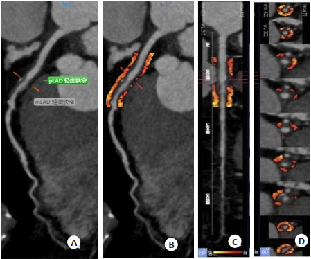
基于双源CT下人工智能CT血流储备分数联合冠状动脉周围脂肪衰减指数对高海拔地区冠状动脉粥样硬化心脏病的评估及预测
doi: 10.12122/j.issn.1674-4500.2022.02.25
Evaluation and prediction of coronary atherosclerotic heart disease in high altitude area by artificial intelligence machine learning-based coronary CT fractional flow reserve combined with coronary perivascular fat attenuation index
-
摘要:
目的 分析基于双源CT下人工智能CT血流储备分数(FFR)联合冠状动脉周围脂肪衰减指数(FAI)对高海拔地区冠状动脉粥样硬化心脏病的评估及预测。 方法 选取2021年10~12月于我院行冠脉CT血管造影(CTA)检查的的63例疑似冠心病患者,以冠脉CTA结果将患者分为血管狭窄率≥50%组与血管狭窄率 < 50%组。比较冠脉狭窄程度、CT-FFR与冠脉周围FAI检查结果;根据冠脉血管钙化程度分组,比较轻中度钙化组、重度钙化组中血管狭窄率≥50%组与 < 50%组的CT-FFR、冠脉周围FAI检查结果,分析各组间CT-FFR与冠脉周围FAI单独与联合的诊断效能。 结果 本研究63例患者中共检查79支冠脉血管,血管狭窄率≥50%组共24支血管,血管狭窄率 < 50%组55支。血管狭窄率≥50%组与血管狭窄率 < 50%组的病变长度差异无统计学意义(P > 0.05),单支血管冠脉狭窄率的差异具有统计学意义(P < 0.05)。血管狭窄率≥50%组的CT-FFR低于血管狭窄率 < 50%组,冠脉周围FAI高于血管狭窄率 < 50%组(P < 0.05)。63例患者中,冠脉血管轻中度钙化47例,重度钙化16例。轻中度钙化组中血管狭窄率≥50%组冠脉狭窄率高于血管狭窄率 < 50%组,CT-FFR低于血管狭窄率 < 50%组(P < 0.05)。重度钙化组中血管狭窄率≥ 50%组CT-FFR低于血管狭窄率 < 50%组,冠脉周围FAI高于血管狭窄率 < 50%组,两组冠脉狭窄率差异无统计学意义(P > 0.05)。ROC曲线显示,CT-FFR+冠脉周围FAI对血管钙化的诊断效能相对于两者单独检查有显著提升(P < 0.05)。 结论 CT-FFR对重度钙化血管诊断效能较低,联合冠脉周围FAI能为血流异常的冠心病患者提高诊断价值。 -
关键词:
- 冠状动脉粥样硬化心脏病 /
- 血流储备分数 /
- 冠脉周围脂肪衰减指数 /
- CT血管造影
Abstract:Objective To investigate the evaluation and prediction of coronary atherosclerotic heart disease in high altitude area by artificial intelligence machine learning-based coronary CT fractional flow reserve (CT-FFR) combined with coronary perivascular fat attenuation index (FAI). Methods Sixty-three patients with suspected coronary artery disease who underwent coronary CT angiography (CTA) in our hospital from October to December 2021 were selected, and the patients were divided into stenosis rate ≥50% group and stenosis rate < 50% group using coronary CTA as the gold standard. The degree of coronary stenosis, CT-FFR and perivascular FAI were compared. Patients were re-divided into two groups according to the degree of coronary artery calcification, mild-to-moderate calcification group and severe calcification group. Then the diagnostic efficacy of CT-FFR and perivascular FAI alone and in combination for abnormal coronary blood flow was evaluated. Results A total of 79 coronary vessels were examined in 63 patients, of which 24 vessels with stenosis rate ≥50% and 55 vessels with stenosis rate < 50%. The lesion length yielded no statistical difference between stenosis rate ≥50% group and stenosis rate < 50% group (P > 0.05), while the proportion of patients with single-vessel coronary stenosis showed statistical difference between two groups (P < 0.05). The CT-FFR in stenosis rate ≥50% group was lower than that in stenosis rate < 50% group, and the perivascular FAI was higher than that in stenosis rate < 50% group (P < 0.05). Among the 63 patients, 47 had mild-to-moderate calcification and 16 had severe calcification. Within mild-to-moderate calcification group, the coronary stenosis rate in stenosis rate ≥50% group was higher than that in stenosis rate < 50% group, and CT-FFR was lower than that in stenosis rate < 50% group. Within severe calcification group, the CT-FFR in stenosis rate ≥50% group was lower than that in stenosis rate < 50% group, and perivascular FAI was higher than that in stenosis rate < 50% group, and no significant difference was found in coronary stenosis rate between stenosis rate < 50% group and stenosis rate ≥50% group (P > 0.05). ROC curve showed that the diagnostic efficiency of CT-FFR+ perivascular FAI for vascular calcification was significantly improved compared with two separate examinations (P < 0.05). Conclusion CT-FFR has low diagnostic efficiency for severe calcification of patients with coronary heart disease, while its combination with perivascular FAI examination achieves high diagnostic value in coronary heart disease patients with blood flow abnormalities. -
表 1 不同FFR组冠脉血管影像学特征
Table 1. Imaging features of coronary vessels in different FFR groups (Mean±SD)
组別 支数 病变长度(mm) 冠脉狭窄率(%) CT-FFR 冠脉周围FAI(Hu) 血管狭窄率≥50%组 24 27.81±12.80 63.84±9.92 0.75±0.03 -73.35±9.52 血管狭窄率 < 50%组 35 23.52±9.54 57.61±9.46 0.87±0.05 -80.62±7.58 t 1.651 2.653 10.908 3.621 P 0.103 0.010 < 0.001 0.001 FFR: 血流储备分数; FAI: 脂肪衰减指数. 表 2 冠脉钙化不同程度血管影像学特征
Table 2. Angiographic features of different degrees of coronary calcification (Mean±SD)
组別 支数 病变长度(mm) 冠脉狭窄率(%) CT-FFR 冠脉周围FAI(Hu) 轻中度钙化 血管狭窄率≥50%组 14 24.83±10.81 64.95±10.68 0.75±0.04 -75.68±9.77 血管狭窄率 < 50%组 45 21.90±8.83 57.21±9.63 0.88±0.06 -81.06±7.04 t 1.027 2.560 7.576 2.269 P 0.309 0.013 < 0.001 0.027 重度钙化 血管狭窄率≥50%组 10 32.15±14.72 62.09±8.87 0.76±0.03 -69.74±8.17 血管狭窄率 < 50%组 10 30.79±9.48 59.70±9.02 0.84±0.06 -82.76±9.31 t 0.246 0.597 3.771 3.324 P 0.809 0.558 0.001 0.004 表 3 CT-FFR与冠脉周围FAI诊断效能
Table 3. Diagnostic efficacy of CT-FFR and pericoronary FAI
方法 AUC 敏感度(%) 特异性(%) P 95%CI 轻中度钙化 CT-FFR 0.763 88.9 57.1 0.003 0.610~0.917 冠脉周围FAI 0.656 95.6 42.9 0.081 0.472~0.839 CT-FFR+冠脉周围FAI 0.790 91.1 57.1 0.001 0.651~0.930 重度钙化 CT-FFR 0.700 80.0 60.0 0.131 0.461~0.939 冠脉周围FAI 0.705 90.0 40.0 0.121 0.473~0.937 CT-FFR+冠脉周围FAI 0.805 90.0 40.0 0.021 0.614~0.996 AUC:曲线下面积. -
[1] Sørensen JK, Framke E, Madsen IEH, et al. 1067Annual changes in job strain and risk of coronary heart disease in Denmark[J]. Int J Epidemiol, 2021, 50(Supplement_1): 202. [2] 买超平, 阎春生, 哈小琴. 海拔高度对冠心病患者EPCs数量及功能的研究[J]. 中国循证心血管医学杂志, 2015, 7(6): 754-7. doi: 10.3969/j.issn.1674-4055.2015.06.09 [3] 杨阿应, 白亚娜, 解芝洞, 等. 2001-2013年金昌队列人群冠心病死亡趋势及疾病负担研究[J]. 中华疾病控制杂志, 2017, 21(3): 261-4, 269. https://www.cnki.com.cn/Article/CJFDTOTAL-JBKZ201703011.htm [4] 靳文剑. 冠状动脉CT血管造影在冠状动脉起源变异中的应用[J]. 中国药物与临床, 2019, 19(7): 1144-5. https://www.cnki.com.cn/Article/CJFDTOTAL-YWLC201907065.htm [5] 李俊灏, 唐春香, 刘通源, 等. 冠状动脉周围脂肪密度指数与斑块参数及血流储备分数关系分析[J]. 中华医学杂志, 2021, 101(39): 3214-20. doi: 10.3760/cma.j.cn112137-20210414-00889 [6] 池黎彤, 刘挨师. CT-FFR对冠状动脉狭窄功能评价的临床价值[J]. 国际医学放射学杂志, 2016, 39(3): 250-3. https://www.cnki.com.cn/Article/CJFDTOTAL-GWLC201603009.htm [7] 陶青, 王胜, 徐峰, 等. 基于CT平扫冠状动脉周围脂肪影像组学诊断非钙化斑块的可行性[J]. 中华医学杂志, 2021, 101(7): 458-63. doi: 10.3760/cma.j.cn112137-20201214-03355 [8] 南丽虹, 李睿君, 冯进堂, 等. 基于CT血管成像的斑块定量分析在冠状动脉血流动力学异常诊断中的应用价值[J]. 中华解剖与临床杂志, 2021, 26(5): 504-10. doi: 10.3760/cma.j.cn101202-20210113-00014 [9] 杨泉, 杨勇, 余建群, 等. 冠状动脉CT造影狭窄程度预测患者远期预后的临床价值研究[J]. 中国全科医学, 2020, 23(12): 1492-6, 1503. doi: 10.12114/j.issn.1007-9572.2019.00.656 [10] 罗发菊. 美国心脏协会[J]. 中华灾害救援医学, 2015, 3(6): 295. https://www.cnki.com.cn/Article/CJFDTOTAL-JYZH201506003.htm [11] 丁熠璞, 单冬凯, 王玺, 等. 冠状动脉周围FAI对CT-FFR诊断重度钙化患者冠脉血流动力学异常的增量价值[J]. 解放军医学杂志, 2021, 46(7): 666-72. https://www.cnki.com.cn/Article/CJFDTOTAL-JFJY202107005.htm [12] Gronewold J, Engels M, van de Velde S, et al. Effects of life events and social isolation on stroke and coronary heart disease[J]. Stroke, 2021, 52(2): 735-47. doi: 10.1161/STROKEAHA.120.032070 [13] 刘明哲. 2型糖尿病并发冠状动脉粥样硬化性心脏病危险因素Logistic回归分析[J]. 分子影像学杂志, 2016, 39(2): 129-33. doi: 10.3969/j.issn.1674-4500.2016.02.18 [14] 张婷婷, 张亚萍. 高海拔地区冠心病患者促甲状腺激素水平与冠状动脉病变程度的关系[J]. 中国循证心血管医学杂志, 2018, 10(2): 176- 8. doi: 10.3969/j.issn.1674-4055.2018.02.13 [15] 刘红, 王引利, 郭良敏, 等. 高海拔贫困地区动脉粥样硬化患者血尿酸水平与饮食习惯的相关性分析[J]. 成都医学院学报, 2019, 14(6): 769-72, 777. doi: 10.3969/j.issn.1674-2257.2019.06.017 [16] 王萍, 周成礼. 颈动脉超声相关指标及动态动脉硬化指数在冠心病风险预测中的意义[J]. 分子影像学杂志, 2017, 40(3): 284-7. doi: 10.3969/j.issn.1674-4500.2017.03.10 [17] 周琦, 王其涛, 蔡芹芹, 等. 冠心病患者左室心尖形态及功能与斑块易损性显著相关[J]. 分子影像学杂志, 2021, 44(2): 332-5. doi: 10.12122/j.issn.1674-4500.2021.02.23 [18] 庞智英, 杨飞, 苏亚英, 等. 冠状动脉CT血管成像联合基于CT的血流储备分数预测阻塞性冠心病主要不良心脏事件的价值[J]. 实用医学杂志, 2021, 37(20): 2675-80. doi: 10.3969/j.issn.1006-5725.2021.20.021 [19] 陈灿, 陶青, 陈蒙, 等. 基于冠状动脉CT钙化积分图像与血管成像测量冠状动脉周围脂肪衰减指数的对比研究[J]. 中华放射学杂志, 2022, 56(3): 254-8. [20] 龚艳君, 易铁慈, 杨帆, 等. 基于冠状动脉CT血管造影的血流储备分数评价心肌缺血的价值[J]. 中国介入心脏病学杂志, 2019, 27(12): 673-8. doi: 10.3969/j.issn.1004-8812.2019.12.002 [21] 李建宜, 许琰, 王俊鹏, 等. 冠状动脉CT血管造影在冠心病患者斑块定量评估及预后评估中的应用价值[J]. 实用临床医药杂志, 2019, 23 (19): 30-2, 36. https://www.cnki.com.cn/Article/CJFDTOTAL-XYZL201919008.htm [22] 何泽兵, 严高武, 李勇, 等. 冠状动脉CT血管造影对先天性右冠状动脉缺如的评价价值[J]. 分子影像学杂志, 2020, 43(4): 606-9. doi: 10.12122/j.issn.1674-4500.2020.04.11 -


 下载:
下载:


