


Comparation of low-dose cardiac scan and coronary CT angiography in quantitative of epicardial adipose tissue volume
-
摘要:
目的 分析低剂量心脏平扫检查与冠状动脉CT血管造影(CTA)检查在定量心外膜脂肪体积(EATV)的一致程度。 方法 对临床提示有冠心病征象的208名患者进行低剂量心脏平扫检查及冠状动脉CTA检查,分别在两种检查的图像上测量心外膜脂肪体积,两种检查测量所得心外膜脂肪体积EATV 心脏平扫和EATVCTA间的相关性采用Pearson相关系数进行评估,采用BlandAltman分析法评价两者的一致性。将患者分为无斑块组及斑块组,比较两组患者EATV 心脏平扫和EATVCTA差异;通过操作者工作特征曲线评估EATV 心脏平扫和EATVCTA预测冠状动脉硬化的价值及差异。 结果 低剂量EATV 心脏平扫为103.12±44.01 cm3,EATVCTA为106.72±44.14 cm3,两者具有显著的相关性(r = 0.955,P< 0.001);EATV 心脏平扫和EATVCTA的一致性均数为-3.5,4.8%(10/208)的点落位于95%CI界限以外(95%CI:24.0930~29.2116,P< 0.001);EATV 心脏平扫和EATVCTA预测冠状动脉硬化发生的曲线下面积分别为0.601及0.605,差异无统计学意义(P= 0.935)。 结论 低剂量心脏平扫检查与冠状动脉CTA两组检查方法在测量EAT方面具有等效性,对冠状动脉硬化的预测方面两者具有相当的价值。 Abstract:Objective To study the consistency between low-dose cardiac plain scanning and coronary artery CT angiography (CTA) in quantification of epicardial adipose tissue volume (EATV). Methods A total of 208 patients with clinically suggestive signs of coronary heart disease were examined by low-dose cardiac plain scan and coronary artery CTA. Epicardial fat volume was measured on the images of both examinations respectively. The correlation between epicardial fat volume measured by the two examinations EATVcardiac plain scan and EATVCTA was evaluated by Pearson correlation coefficient. Bland-altman analysis was used to evaluate their consistency. Patients were divided into the plaque free group and the plaque groups, and the differences of EATVcardiac plain scan and EATVCTA between the two groups were compared. ROC curve was used to evaluate the value and difference of EATVcardiac plain scan and EATVCTA in predicting coronary atherosclerosis. Results There was a significant correlation between low-dose EATV cardiac sweep of 103.12±44.01 cm3 and EATVCTA of 106.72±44.14 cm3 (r = 0.955, P< 0.001). The mean consistency between EATVcardiac plain scan and EATVCTA was -3.5, with 4.8% (10/208) points falling outside the 95%CI (95%CI: 24.0930- 29.2116, P< 0.001). The area under the curve for predicting by EATVcardiac plain scan and EATVCTA were 0.601 and 0.605 respectively, with no statistical difference between the two groups (P= 0.935). Conclusion Low-dose cardiac plain scan and coronary artery CTA are equivalent in the measurement of EAT, and both are of equal value in the prediction of coronary atherosclerosis. -
Key words:
- epicardial adipose tissue volume /
- low dose /
- plain cardiac scan /
- tomography
-

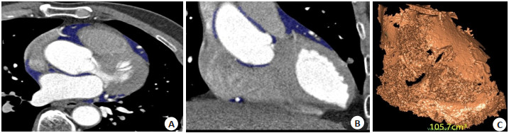
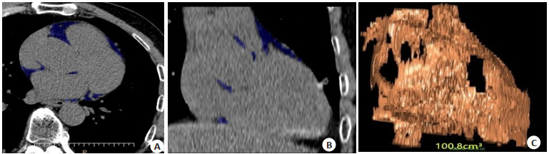
图 1 EAT心脏平扫心外膜脂肪组织分割
A:心脏平扫轴位重组图; B:心脏平扫矢状位重组图; C:心脏平扫体积计算图.
Figure 1. Epicardial adipose tissue segmentation on cardiac plain scan.

图 2 EAT CTA心外膜脂肪组织分割
A:心脏CTA轴位重组图; B:心脏CTA矢状位重组图; C:心脏CTA体积计算图.
Figure 2. Epicardial adipose tissue segmentation on CTA.

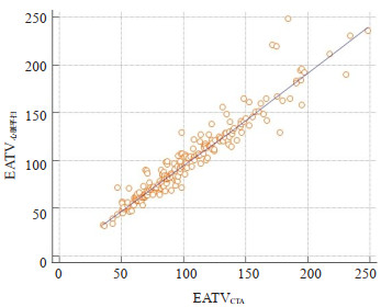
图 3 EATV心脏平扫及EATVCTA两组相关性分析图
Figure 3. Correlation analysis of EATVcardiac plain scan group and EATVCTA group.

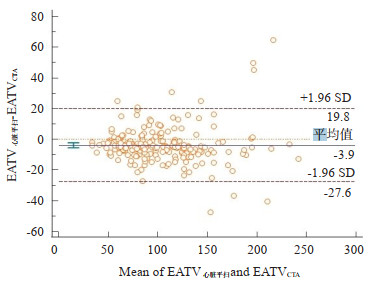
图 4 EATV心脏平扫及EATVCTA两组一致性Bland-Altman图
Figure 4. Consistent Bland- Altman plot of EATVcardiac plain scan group and EATVCTA group.

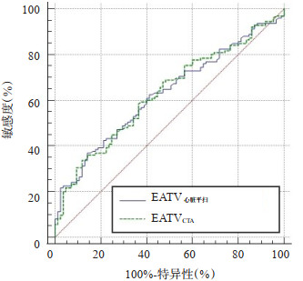
图 5 EATV心脏平扫及EATVCTA两组ROC曲线
Figure 5. ROC curves of EATVcardiac plain scanand EATVCTA.
表 1 EATV心脏平扫及EATVCTA测量在无斑块组与斑块组的差异比较
Table 1. Differences of EATVcardiac plain scan and EATCTA measurements between the non-plaque group and the plaque group (cm3, Mean±SD)
方法 无斑块组(n=85) 斑块组(n=123) t P EATV心脏平扫 92.77±3.55 107.70±3.97 2.658 0.008 EATVCTA 96.17±3.57 111.32±3.86 2.744 0.006  下载: 导出CSV
-
[1] Du Y, Yang L, Liu Y, et al. Relation between quantity and quality of peri-coronary epicardial adipose tissue and its underlying hemodynamically significant coronary Stenosis[J]. BMC Cardiovasc Disord, 2020, 20(1): 226. doi: 10.1186/s12872-020-01499-w [2] Gao Z, Zuo Y, Jia L, et al. Association between epicardial adipose tissue density and characteristics of coronary plaques assessed by coronary computed tomographic angiography[J]. Int J Cardiovasc Imaging, 2021: 37(11): 3177-80. doi: 10.1007/s10554-021-02396-9 [3] Ma RL, van Assen M, Ties D, et al. Focal pericoronary adipose tissue attenuation is related to plaque presence, plaque type, and Stenosis severity in coronary CTA[J]. Eur Radiol, 2021, 31(10): 7251-61. doi: 10.1007/s00330-021-07882-1 [4] 李红文, 赵韧, 李小虎, 等.心外膜脂肪体积与冠状动脉粥样硬化及心肌桥的关系[J].中国医学影像技术, 2020, 36(1): 64-7. https://www.cnki.com.cn/Article/CJFDTOTAL-ZYXX202001023.htm [5] Svanteson M, Holte KB, Haig Y, et al. Coronary plaque characteristics and epicardial fat tissue in long term survivors of type 1 diabetes identified by coronary computed tomography angiography[J]. Cardiovasc Diabetol, 2019, 18(1): 58. doi: 10.1186/s12933-019-0861-x [6] Klein C, Brunereau J, Lacroix D, et al. Left atrial epicardial adipose tissue radiodensity is associated with electrophysiological properties of atrial myocardium in patients with atrial fibrillation[J]. Eur Radiol, 2019, 29(6): 3027-35. doi: 10.1007/s00330-018-5793-4 [7] Hwang EJ, Goo JM, Kim HY, et al. Optimum diameter threshold for lung nodules at baseline lung cancer screening with low-dose chest CT: exploration of results from the Korean Lung Cancer Screening Project[J]. Eur Radiol, 2021, 31(9): 7202-12. doi: 10.1007/s00330-021-07827-8 [8] Erkmen CP, Dako F, Moore R, et al. Adherence to annual lung cancer screening with low-dose CT scan in a diverse population[J]. Cancer Causes Control, 2021, 32(3): 291-8. doi: 10.1007/s10552-020-01383-0 [9] Nagayama Y, Nakamura N, Itatani R, et al. Epicardial fat volume measured on nongated chest CT is a predictor of coronary artery disease[J]. Eur Radiol, 2019, 29(7): 3638-46. doi: 10.1007/s00330-019-06079-x [10] Lee KC, Yong HS, Lee J, et al. Is the epicardial adipose tissue area on non-ECG gated low-dose chest CT useful for predicting coronary atherosclerosis in an asymptomatic population considered for lung cancer screening?[J]. Eur Radiol, 2019, 29(2): 932-40. doi: 10.1007/s00330-018-5562-4 [11] Yang JJ, Dou GH, Tesche C, et al. Progression of coronary atherosclerotic plaque burden and relationship with adverse cardiovascular event in asymptomatic diabetic patients[J]. BMC Cardiovasc Disord, 2019, 19(1): 39. doi: 10.1186/s12872-019-1016-4 [12] 程凯, 查云飞, 胡磊, 等.心脏CT平扫与CTA定量分析心外膜脂肪体积对比研究[J]. CT理论与应用研究, 2019, 28(5): 585-91. https://www.cnki.com.cn/Article/CJFDTOTAL-CTLL201905013.htm [13] Ma RL, Ties D, van Assen M, et al. Towards reference values of pericoronary adipose tissue attenuation: impact of coronary artery and tube voltage in coronary computed tomography angiography [J]. Eur Radiol, 2020, 30(12): 6838-46. doi: 10.1007/s00330-020-07069-0 [14] Yan CG, Liang CY, Xu J, et al. Ultralow-dose CT with knowledgebased iterative model reconstruction (IMR) in evaluation of pulmonary tuberculosis: comparison of radiation dose and image quality[J]. Eur Radiol, 2019, 29(10): 5358-66. doi: 10.1007/s00330-019-06129-4 [15] Chamberlin J, Kocher MR, Waltz J, et al. Automated detection of lung nodules and coronary artery calcium using artificial intelligence on low-dose CT scans for lung cancer screening: accuracy and prognostic value[J]. BMC Med, 2021, 19(1): 55. doi: 10.1186/s12916-021-01928-3 [16] Zhu YQ, Wang Y, Gioia WE, et al. Visual scoring of aortic valve calcifications on low-dose CT in lung cancer screening[J]. Eur Radiol, 2020, 30(5): 2658-68. doi: 10.1007/s00330-019-06614-w [17] Gać P, Macek P, Poręba M, et al. Thickness of epicardial and pericoronary adipose tissue measured using 128-slice MSCT as predictors for risk of significant coronary artery diseases[J]. Ir J Med Sci, 2021,190(2): 555-66. doi: 10.1007/s11845-020-02339-8 [18] Yu W, Zhang F, Liu B, et al. Incremental value of epicardial fat volume to coronary artery calcium score and traditional risk factors for predicting myocardial ischemia in patients with suspected coronary artery disease[J]. J Nucl Cardiol, 2021: 2021Feb19. [19] la Grutta L, Toia P, Farruggia A, et al. Quantification of epicardial adipose tissue in coronary calcium score and CT coronary angiography image data sets: comparison of attenuation values, thickness and volumes[J]. Br J Radiol, 2016, 89(1062): 20150773. doi: 10.1259/bjr.20150773 [20] Lim C, Ahn MI, Jung JI, et al. Simple quantification of paracardial and epicardial fat dimensions at low-dose chest CT: correlation with metabolic risk factors and usefulness in predicting metabolic syndrome[J]. Jpn J Radiol, 2018, 36(9): 528-36. doi: 10.1007/s11604-018-0752-1 [21] Zhang L, Sun JQ, Jiang BB, et al. Development of artificial intelligence in epicardial and pericoronary adipose tissue imaging: a systematic review[J]. Eur J Hybrid Imaging, 2021, 5: 14. doi: 10.1186/s41824-021-00107-0 [22] Slart RHJA, Williams MC, Juarez-Orozco LE, et al. Position paper of the EACVI and EANM on artificial intelligence applications in multimodality cardiovascular imaging using SPECT/CT, PET/CT, and cardiac CT[J]. Eur J Nucl Med Mol Imaging, 2021, 48(5): 1399-413. doi: 10.1007/s00259-021-05341-z -

 点击查看大图
计量
- 文章访问数: 242
- HTML全文浏览量: 117
- PDF下载量: 13
- 被引次数: 0