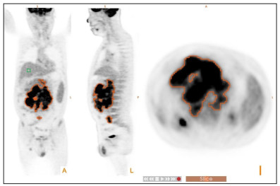
18F-FDG PET/CT参数SUVpeak、全身代谢肿瘤体积和总糖酵解值对弥漫大B细胞淋巴瘤患者预后的预测价值
doi: 10.12122/j.issn.1674-4500.2021.05.11
Predictive value of 18F-FDG PET/CT quantization parameters SUVpeak, MTV and TLG in patients with diffuse large B-cell lymphoma
-
摘要:
目的 探讨化疗前PET/CT检查的SUVpeak、代谢肿瘤体积(MTV)、总糖酵解值(TLG)是否可以预测弥漫大B细胞淋巴瘤患者的预后。 方法 回顾性分析2011年8月~2018年11月本院诊治的76例弥漫大B细胞淋巴瘤患者资料,根据随访 结果 将患者分为进展组(n=40)和未进展组(n=36)、死亡组(n=6)和存活组(n=70)。所有患者化疗前均接受18F-FDG PET/CT检查,使用自动感兴趣区勾画软件测量记录病灶的SUVmax、SUVpeak、MTV、TLG等参数。比较疾病进展组和未进展组之间、死亡组和存活组之间PET参数的差异,通过ROC曲线求取PET参数的最佳临界值,并采用Kaplan-Meier生存分析比较生存率,使用临床变量资料和PET参数对生存期和疾病进展进行单因素和多因素分析。 结果 疾病进展组的中位SUVmax、SUVpeak、MTV和TLG分别为30.82(12.64~71.16)、19.89(9.35~52.23)、786.49(135.06~8795.16)cm3、4618.76(653.88~37361.18);未进展组为34.53(3.82~52.41)、15.76(3.18~32.37)、116.05(54.14~2642.96)cm3、420.18(276.97~9409.09)。进展组和未进展组之间SUVpeak、MTV及TLG差异有统计学意义(P < 0.05)。死亡组的中位数SUVmax、SUVpeak、MTV和TLG分别为50.47(19.95~130.14)、37.40(16.95~82.63)、2195.11(231.85~8361.82)cm3、14712.77(3371.5~28302.65);存活组为30.82(3.82~52.41)、19.43(3.18~38.62)、252.10(54.14~ 8795.16)cm3,1219.53(276.97~37361.18)。死亡组和存活组之间SUVmax、SUVpeak、MTV及TLG数值的差异均有统计学意义(P < 0.05)。患者SUVpeak、MTV和TLG小于临界值时患者生存率明显高于其大于临界值时。SUVpeak、MTV、TLG及国际预后指数评分与无进展生存期呈显著相关。 结论 SUVpeak、MTV和TLG均能在弥漫大B细胞淋巴瘤患者的治疗预后中提供重要的预测价值。 Abstract:Objective To evaluate the predictive significance of F-18 FDG PET/CT quantization parameters SUVpeak, MTV and TLG in patients with diffuse large B cell lymphoma (DLBCL) before chemotherapy. Methods Seventy-six patients with DLBCL were involved between August 2011 and November 2018 who had undergone F-18 FDG PET/CT scan prior to treatment. The patients were divided into progress group (n=40) and non-progressive group (n=36), death group (n=6) and survival group (n= 70). Maximum standardized uptake value (SUVmax), peak standardized uptake value (SUVpeak), metabolic tumor volume (MTV) and total lesion glycolysis (TLG) were measured by a automatic VOI software. The differences in PET parameters between the progression group and the non-progressive group, and between the death group and the survival group were compared. The best cut-off value of PET parameters through the ROC curve was obtained. Kaplan-Meier survival analysis was used to compare survival rates. The clinical variables Data and PET parameters were used for univariate and multivariate analysis of survival and progression. Methods The median SUVmax, SUVpeak, MTV and TLG in the progression group were 30.82(12.64-71.16), 19.89(9.35-52.23), 786.49(135.06-8795.16) cm3 and 4618.76(653.88-37361.18), and 34.53(3.82-52.41), 15.76(3.18-32.37), 116.05 (54.14-2642.96) cm3 and 420.18(276.97-9409.09) in the non-progressive group. The differences in SUVpeak, MTV and TLG between progressive group and non progressive group were significant(p < 0.05). The median SUVmax, SUVpeak, MTV and TLG were 50.47 (19.95-130.14), 37.40(16.95-82.63), 2195.11(231.85-8361.82) cm3 and 14712.77(3371.5-28302.65) in the death group; and 30.82 (3.82-52.41), 19.43(3.18-38.62), 252.10(54.14-8795.16) cm3 and 1219.53(276.97-37361.18) in the survival group.The differences in the SUVmax, SUVpeak, MTV, and TLG between the dead group and the survival group were significant (P < 0.05). The survival rate of patients when SUVpeak, MTV and TLG are less than the cut-off value was significantly higher than when they are greater than the cut-off value. SUVpeak, MTV, TLG and international prognostic index scores were positively correlated with progression-free survival. Conclusions SUVpeak, MTV and TLG can all provide important predictive value in the prognosis of DLBCL patients. -
Key words:
- PET/CT /
- DLBCL /
- quantization parameters /
- predictive value /
- Progression-Free-Survival
-
表 1 DLBCL患者临床特征
Table 1. Patient characteristics
特征 n (%) 性別 男 54(71.05) 女 22(28.95) Ann Arbor分期 Ⅰ 5(6.58) Ⅱ 8(10.53) Ⅲ 16(21.05) Ⅳ 47(61.84) IPI评分 0~1 15(19.74) 2~5 61(80.26) 亚型 GCB 30 (39.47) non-GCB 46 (60.53) IPI: 国际预后指数;GCB: 生发中心来源; DLBCL: 弥漫大B细胞淋巴瘤 表 2 单变量回归分析
Table 2. Univariate Cox proportional hazard regression analysis
因素 HR 95%CI P SUVmax 1.049 0.976~1.133 0.254 GCB 0.917 0.522~1.612 0.665 Non-GCB 1.101 0.943~1.118 0.398 SUVpeak 1.036 1.018~1.067 0.032 GCB 1.024 1.013~1.047 0.024 Non-GCB 1.045 1.012~1.089 0.040 MTV 1.032 1.014~1.046 < 0.001 GCB 1.028 1.008~1.043 0.016 Non-GCB 1.082 1.056~1.123 0.002 TLG 1.078 1.040~1.082 < 0.001 GCB 1.090 1.031~1.142 0.007 Non-GCB 1.112 1.078~1.182 0.004 IPI 1.580 1.247~2.089 0.005 LDH 1.003 0.988~1.012 0.223 Ki-67 0.814 0.091~6.122 0.782 MTV:代谢肿瘤体积;TLG:总糖酵解值;LDH:乳酸脱氢酶. 表 3 多变量回归分析
Table 3. Multivariate Cox proportional hazard regression analysis
因素 HR 95%CI P SUVmax 1.042 0.982~1.121 0.244 SUVpeak 1.025 1.011~1.046 0.028 MTV 1.064 1.035~1.102 0.001 TLG 1.088 1.040~1.082 0.001 -
[1] 应志涛, 王雪鹃, 宋玉琴, 等. 弥漫大B细胞淋巴瘤患者规范治疗后行18F-FDG PET/CT检查的预后意义[J]. 中华血液学杂志, 2012, 33 (10): 810-3. doi: 10.3760/cma.j.issn.0253-2727.2012.10.005 [2] 中华医学会核医学分会PET与分子影像学组. 淋巴瘤18F-FDG PET/ CT显像临床应用指南(2016版)[J]. 中华核医学与分子影像杂志, 2016, 36(5): 458-60. doi: 10.3760/cma.j.issn.2095-2848.2016.05.017 [3] Juweid ME, Stroobants S, Hoekstra OS, et al. Use of positron emission tomography for response assessment of lymphoma: consensus of the imaging subcommittee of international harmonization project in lymphoma[J]. J Clin Oncol, 2007, 25(5): 571-8. doi: 10.1200/JCO.2006.08.2305 [4] Schot B, van Imhoff G, Pruim J, et al. Predictive value of early 18Ffluoro-deoxyglucose positron emission tomography in chemosensitive relapsed lymphoma[J]. Br J Haematol, 2003, 123 (2): 282-7. doi: 10.1046/j.1365-2141.2003.04593.x [5] 刁玉竹, 洪成雨, 赵英杰, 等. 18F-FDG PET-CT在淋巴瘤中的应用及与弥漫大B细胞淋巴瘤预后的关系[J]. 临床肿瘤学杂志, 2020, 25 (7): 631-5. doi: 10.3969/j.issn.1009-0460.2020.07.010 [6] Sharma P, Gupta A, Patel C, et al. Pediatric lymphoma: metabolic tumor burden as a quantitative index for treatment response evaluation[J]. Ann Nucl Med, 2012, 26(1): 58-66. doi: 10.1007/s12149-011-0539-2 [7] Cheson BD, Pfistner B, Juweid ME, Gascoyne RD, Specht L, Horning SJ, et al. Revised response criteria for malignant lymphoma [j]. J Clin Oncol. 2007;25: 579–86. doi: 10.1200/JCO.2006.09.2403 [8] 苏洁敏, 郑庆中, 黄文荣, 等. 18F-FDG PET/CT代谢参数预测弥漫大B细胞淋巴瘤患者预后的价值[J]. 中国实验血液学杂志, 2021, 29(4): 1181-6. https://www.cnki.com.cn/Article/CJFDTOTAL-XYSY202104027.htm [9] 孙悦, 许宏, 郭振清, 等. 探索18F-FDG PET/CT SUVmax、SUVsum及病理Ki-67表达等在非霍奇金淋巴瘤中的临床应用价值[J]临床血液学杂志, 2021, 34(1): 18-23. https://www.cnki.com.cn/Article/CJFDTOTAL-LCXZ202101005.htm [10] Adams HJA, de Klerk JMH, Fijnheer R, et al. Prognostic superiority of the National Comprehensive Cancer Network International Prognostic Index over pretreatment whole-body volumetricmetabolic FDG-PET/CT metrics in diffuse large B-cell lymphoma [J]. Eur J Haematol, 2015, 94(6): 532-9. doi: 10.1111/ejh.12467 [11] Sasanelli M, Meignan M, Haioun C, et al. Pretherapy metabolic tumour volume is an independent predictor of outcome in patients with diffuse large B-cell lymphoma[J]. Eur J Nucl Med Mol Imaging, 2014, 41(11): 2017-22. doi: 10.1007/s00259-014-2822-7 [12] Zhang YY, Song L, Zhao MX, et al. A better prediction of progression-free survival in diffuse large B-cell lymphoma by a prognostic model consisting of baseline TLG and %ΔSUVmax[J]. Cancer Med, 2019, 8(11): 5137-47. doi: 10.1002/cam4.2284 [13] Islam P, Goldstein JS, Sethi I, et al. PET-derived tumor metrics predict DLBCL response and progression-free survival[J]. Blood, 2017, 130: 4140. http://www.onacademic.com/detail/journal_1000041611585199_7d10.html [14] 赵鹏, 朱博, 赵尉, 等. 肿瘤基线代谢参数预测弥漫大B细胞淋巴瘤预后的价值[J]. 中国医科大学学报, 2019, 48(12): 1122-6. https://www.cnki.com.cn/Article/CJFDTOTAL-ZGYK201912014.htm [15] Ceriani L, Martelli M, Conconi A, et al. Prognostic models for primary mediastinal (thymic) B-cell lymphoma derived from 18- FDG PET/CT quantitative parameters in the International Extranodal Lymphoma Study Group (IELSG) 26 study[J]. Br J Haematol, 2017, 178(4): 588-91. doi: 10.1111/bjh.14728 [16] Boellaard R, Krak N, Hoekstra O, et al. Effects of noise, image resolution, and ROI definition on the accuracy of standard uptake values: a simulation study[J]. J Nucl Med, 2004, 45(9): 1519-27. http://jnm.snmjournals.org/cgi/reprint/45/9/1519.pdf [17] Moon SH, Choi JY, Lee HJ, et al. Prognostic value of 18F-FDG PET/ CT in patients with squamous cell carcinoma of the tonsil: comparisons of volume-based metabolic parameters[J]. Head Neck, 2013, 35(1): 15-22. doi: 10.1002/hed.22904 [18] Xie MX, Zhai WH, Cheng SY, et al. Predictive value of F-18 FDG PET/CT quantization parameters for progression-free survival in patients with diffuse large B-cell lymphoma[J]. Hematology, 2016, 21(2): 99-105. doi: 10.1179/1607845415Y.0000000033 [19] Esfahani SA, Heidari P, Halpern EF, et al. Baseline total lesion glycolysis measured with (18)F-FDG PET/CT as a predictor of progression-free survival in diffuse large B-cell lymphoma: a pilot study[J]. Am J Nucl Med Mol Imaging, 2013, 3(3): 272-81. http://europepmc.org/articles/PMC3627523 [20] Gallicchio R, Mansueto G, Simeon V, et al. F-18 FDG PET/CT quantization parameters as predictors of outcome in patients with diffuse large B-cell lymphoma[J]. Eur J Haematol, 2014, 92(5): 382-9. doi: 10.1111/ejh.12268 -


 下载:
下载:


