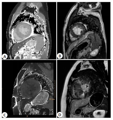
Application of gadolinium delayed enhancement combined with cardiac magnetic resonance T1 mapping in cardic amyloidosis and hypertrophic cardiomyopathy: prospective study of 95 patients
-
摘要:
目的 研究钆延迟强化联合心脏磁共振T1 mapping在心肌淀粉样变性(CA)和肥厚性心肌病(HCM)患者临床检查中的应用。 方法 采用前瞻性研究方法,以我院2018年12月~2020年12月诊断的CA患者40例和HCM患者55例作为研究对象,另选取同期进行健康体检的患者54例作为对照组,比较3组患者的心功能、心脏形态学、MRI相关指标之间的差异。 结果 左心室收缩期末容积、左心室射血分数在3组间的差异存在统计学意义(P < 0.05),通过两两比较,左心室收缩期末容积、左心室射血分数从高到低依次为HCM组、对照组、CA组,3组患者的LVEF差异不存在统计学意义(P > 0.05);左室壁厚度以及左心室质量从高到低依次为HCM组、CA组以及对照组(P < 0.05);增强前T1值从高到低依次为CA组、HCM组以及对照组(P < 0.05);增强后T1值从高到低依次为HCM组、对照组以及CA组(P < 0.05);细胞外容积从高到低依次为CA组、对照组以及HCM组(P < 0.05)。 结论 钆延迟强化联合心脏磁共振T1 mapping在CA和HCM患者临床检查中具有较好的鉴别诊断能力,建议临床推广。 -
关键词:
- 钆延迟强化 /
- 心脏磁共振 /
- T1 mapping /
- 联合检测
Abstract:Objective To explore the application of gadolinium delayed enhancement combined with cardiac magnetic resonance T1 mapping in the clinical examination of patients with cardic amyloidosis (CA) and hypertrophic cardiomyopathy (HCM). Methods In this prospective study, 40 CA patients and 55 HCM patients diagnosed in our hospital from December 2018 to December 2020 were selected as the research objects, and 54 healthy patients were selected as the control group. The cardiac function, cardiac morphology, and MRI related indicators in three groups were compared. Results The difference between the levels of LVESV and LVEDV of three groups was significant (P < 0.05). By pairwise comparison, the levels of LVESV and LVEDV were the HCM group, the control group and the CA group in descending order. There was no significant difference in LVEF between the three groups of patients (P > 0.05). The left ventricular wall thickness and left ventricular mass of the three groups were the HCM group, the CA group and the control group in descending order (P < 0.05). The T1 values of the three groups were the CA Group, the HCM group and the control group in descending order (P < 0.05). After the enhancement, the T1 value was the HCM group, the control group and the CA group in descending order. The extracellular volume was the CA group, the control group and the HCM group in descending order (P < 0.05). Conclusion Gadolinium delayed enhancement combined with cardiac magnetic resonance T1 mapping has a good differential diagnosis ability in the clinical examination of CA and HCM patients. -
表 1 一般资料比较
Table 1. Comparison of general information
项目 性別(男/女,n) 年龄(岁,Mean±SD) BMI(kg/m2, Mean±SD) CA组(n=40) 16/24 46.29±2.05 24.55±3.43 HCM组(n=55) 22/33 46.55±2.42 24.30±3.09 对照组(n=54) 21/33 46.41±2.79 24.69±3.44 F/χ2 1.427 0.898 0.593 P 0.426 0.370 0.554 CA: 心肌淀粉样变性; HCM: 肥厚性心肌病. 表 2 心功能比较
Table 2. Comparison of cardiac function (Mean±SD)
项目 LVESV(mL/m2) LVEDV(mL/m2) LVEF(%) CA组(n=40) 49.74±11.37 85.21±21.95 56.33±11.33 HCM组(n=55) 69.70±11.51 141.60±21.96 51.37±11.57 对照组(n=54) 50.65±11.45 119.21±21.77 53.10±11.77 F 11.236 10.239 0.698 P < 0.001 < 0.001 0.336 LSD-t(CA vs HCM) 10.336 9.887 - P < 0.001 < 0.001 - LSD-t(CA vs对照) 9.584 9.996 - P < 0.001 < 0.001 - LSD-t(对照vs HCM) 8.256 7.559 - P < 0.001 < 0.001 - 表 3 三组研究对象的心脏形态学指标比较
Table 3. Comparison of cardiac morphological indexes among three groups (Mean±SD)
项目 左室壁厚度(mm) 左心室质量(g) CA组(n=40) 13.96±2.63 160.52±2.14 HCM组(n=55) 16.99±1.96 211.03±12.14 对照组(n=54) 6.91±2.47 105.32±1.45 F 11.230 12.698 P < 0.001 < 0.001 LSD-t(CA vs HCM) 9.885 9.647 P < 0.001 < 0.001 LSD-t(CA vs对照) 8.552 8.698 P < 0.001 < 0.001 LSD-t(对照vs HCM) 7.885 6.997 P < 0.001 < 0.001 表 4 核磁共振的相关指标比较
Table 4. Comparison of MRI related indexes(Mean±SD)
项目 增强前T1值 增强后T1值 细胞外容积(%) CA组(n=40) 1481.77±87.24 455.27±3.31 48.04±4.24 HCM组(n=55) 1381.27±66.94 578.77±3.27 26.88±4.17 对照组(n=54) 1273.77±77.24 521.27±3.31 27.04±4.24 F 12.369 15.698 15.741 P < 0.001 < 0.001 < 0.001 LSD-t(CA vs HCM) 10.258 10.697 11.587 P < 0.001 < 0.001 < 0.001 LSD-t(CA vs对照) 9.885 10.587 10.555 P < 0.001 < 0.001 < 0.001 LSD-t(对照vs HCM) 9.885 10.202 10.636 P < 0.001 < 0.001 < 0.001 -
[1] 崔倩, 于静, 沈文. 钆延迟强化联合纵向弛豫时间定量成像评估心肌淀粉样变性的价值[J]. 中华危重病急救医学, 2019, 31 (12): 1538-41. doi: 10.3760/cma.j.issn.2095-4352.2019.12.021 [2] 冀晋, 方理刚, 方全, 等. 超声心动图二维斑点追踪成像与心脏核磁共振钆延迟增强对心肌淀粉样变性检测的比较[J]. 中国循环杂志, 2018, 33(1): 87-91. doi: 10.3969/j.issn.1000-3614.2018.01.017 [3] 王立明, 田颖, 赵蕾, 等. 免疫球蛋白轻链型心肌淀粉样变性临床分析[J]. 中国全科医学, 2020, 23(27): 3474-8. doi: 10.12114/j.issn.1007-9572.2020.00.048 [4] 朱强, 胡信群, 唐亮, 等. 心肌淀粉样变性一例[J]. 中国循环杂志, 2017, 32(2): 122. [5] Narotsky DL, Castano A, Weinsaft JW, et al. Wild-type transthyretin cardiac amyloidosis: novel insights from advanced imaging[J]. Can J Cardiol, 2016, 32(9): . [6] De Bruijn S, Galloo X, De Keulenaer G, et al. A special case of hypertrophic cardiomyopathy with a differential diagnosis of isolated cardiac amyloidosis or junctophilin type 2 associated cardiomyopathy[J]. Acta Clin Belg, 2021, 76(2): 136-43. doi: 10.1080/17843286.2019.1662572 [7] Wang S, Wang QL, Zhai N, et al. Progression of Danon disease with medical imaging: two case reports[J]. J Int Med Res, 2021, 49 (2): 030006052098667. [8] Pulsford C. Anaesthetic management of underlying hypertrophic cardiomyopathy in the feline dental patient: an extended patient care report[J]. Vet Nurs J, 2021, 36(2): 55-9. doi: 10.1080/17415349.2021.1876538 [9] Masri A, Nazer B, Al-Rashdan L, et al. Thirty controversies and considerations in hypertrophic cardiomyopathy[J]. Struct Heart, 2021, 5(1): 39-54. doi: 10.1080/24748706.2020.1844926 [10] Natarajan R, Ameduri R, Griselli M, et al. Angiographic evidence of backward compression wave: systolic compression of septal perforators in a child with hypertrophic cardiomyopathy[J]. Cardiol Young, 2021, 31(1): 125-6. doi: 10.1017/S104795112000428X [11] Rujirachun P, Charoenngam N, Wattanachayakul P, et al. Efficacy and safety of direct oral anticoagulants (DOACs) versus vitamin K antagonist (VKA) among patients with atrial fibrillation and hypertrophic cardiomyopathy: a systematic review and meta-analysis[J]. Acta Cardiol, 2020, 75(8): 724-31. doi: 10.1080/00015385.2019.1668113 [12] Coats AJ. Future developments in the MECKI score initiative[J]. Eur J Prev Cardiol, 2020, 27(2_suppl): 72-5. [13] Briller J, Vaught AJ. Cardiomyopathies in pregnancy: etiologies and management strategies[J]. Clin Obstet Gynecol, 2020, 63(4): 893-909. doi: 10.1097/GRF.0000000000000568 [14] Darwish RK, Haghighi A, Seliem ZS, et al. Genetic study of pediatric hypertrophic cardiomyopathy in Egypt[J]. Cardiol Young, 2020, 30(12): 1910-6. doi: 10.1017/S1047951120003157 [15] Siegrist KK, Deegan RJ, Dumas SD, et al. Severe cardiopulmonary disease in a parturient with noonan syndrome[J]. Semin Cardiothorac Vasc Anesth, 2020, 24(4): 364-8. doi: 10.1177/1089253220945918 [16] 张义, 王万虹, 崔文, 等. 3.0T磁共振延迟强化技术检测存活心肌评估CTO-PCI术后心功能恢复情况[J]. 中国临床研究, 2021, 34(3): 299-303. [17] 李文霞, 王静, 杨帆, 等. 心电图和超声心动图对肥厚型心肌病钆延迟强化的预测研究[J]. 中华超声影像学杂志, 2018, 27(8): 645-9. doi: 10.3760/cma.j.issn.1004-4477.2018.08.001 [18] 谢亚闯, 谌小丽, 董新博. 心脏磁共振钆对比剂延迟强化对扩张型心肌病患者心脏不良事件的预测价值研究[J]. 实用心脑肺血管病杂志, 2017, 25(6): 9-13. doi: 10.3969/j.issn.1008-5971.2017.06.003 [19] 马义泼, 黄健, 盛二燕, 等. 心脏磁共振钆对比剂延迟强化的临床意义及判断预后的预测价值[J]. 影像研究与医学应用, 2019, 3 (17): 24-6. [20] 纳丽莎, 刘蕾, 朱力, 等. 超声三维斑点追踪成像联合心脏磁共振成像-钆延迟增强序列对成人肥厚型心肌病左室整体收缩功能与心肌纤维化的关联性研究[J]. 中华超声影像学杂志, 2020, 29 (1): 6-12. DOI: 10.3760/cma.j.issn.1004-4477.2020.01.002. [21] 宋德利, 郑帅, 刘彤, 等. 心脏磁共振钆延迟强化程度与慢性心力衰竭患者预后的关系[J]. 心肺血管病杂志, 2017, 36(8): 692-5, 700. doi: 10.3969/j.issn.1007-5062.2017.08.021 [22] 曾道兵, 赵新湘, 常婵. 钆延迟增强心脏磁共振识别的隐性心肌梗死特征及其与冠状动脉狭窄的相关性[J]. 中国循环杂志, 2019, 34(3): 234-8. doi: 10.3969/j.issn.1000-3614.2019.03.005 [23] 张青, 王振常, 赵波, 等. 心脏磁共振延迟强化成像定量评价心肌梗死的研究[J]. 临床荟萃, 2007, 22(10): 696-9. doi: 10.3969/j.issn.1004-583X.2007.10.004 [24] 张晓, 赵猛, 王静. 心肌淀粉样变性MRI及临床特征分析(附4例报道[) J]. 医学影像学杂志, 2020, 30(6): 981-3. [25] 朱孔博, 程中伟, 田庄, 等. 心脏核磁共振在心肌淀粉样变中的诊断价值[J]. 中华心血管病杂志, 2011, 39(10): 915-9. doi: 10.3760/cma.j.issn.0253-3758.2011.10.009 [26] 张学强, 相世峰, 杨素君, 等. 心脏磁共振成像在左心室肥厚病变鉴别诊断中的价值[J]. 医学影像学杂志, 2018, 28(6): 920-4. -


 下载:
下载:


