


Pneumonia X-ray images enhance classification recognition technology: Based on improved Retinex algorithm
-
摘要:
目的 引入并改进Retinex算法增强肺图像特征,提升肺炎识别的准确率。 方法 提出一种对肺炎X光图像特征增强的Retinex优化方法。将X光图边缘中心化,进行信息重建,利用Retinex进行特征强化,最后将图像赋予权重与原图像相结合,最大程度保留特征。 结果 相对于初始图像训练,其准确率提升了2.57个百分点,损失了0.17个百分点的敏感度准确率,却增加了7.15个百分点的特异性准确率。 结论 改进后的算法能够使得机器快速自动识别肺炎与非肺炎,在肺炎高发的时期大大提升了诊断效率。 Abstract:Objective To enhance Retinex optimization method and pneumonia X- ray image features. Methods A Retinex optimization method for enhancement of pneumonia X-ray image features was proposed. The edge of the X-ray image was centralized for information reconstruction. Retinex was used for feature enhancement. The image was weighted and combined with the original image to retain features to the maximum extent. Results Compared with the initial image training, the accuracy was increased by 2.57 percentage points, the sensitivity accuracy was lost by 0.17 percentage points, and the specificity accuracy was increased by 7.15 percentage points. Conclusion The improved algorithm can enable the machine to quickly and automatically identify pneumonia and non-pneumonia, which greatly improves the diagnostic efficiency during the period of high incidence of pneumonia. -
Key words:
- deep learning /
- pneumonia classification /
- image processing /
- retinex algorithm
-

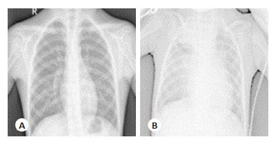
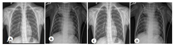
图 1 图像重建对比结果
A:正常肺部初始图像;B:患肺炎肺部初始图像;C:正常肺部重建图像;D:患肺炎肺部重建图像.
Figure 1. Image reconstruction contrast results.

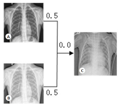
图 3 Retinex算法特征强化结果
A:肺部重建强化图像;B:肺炎重建强化图像.
Figure 3. Retinex algorithm feature-enhanced results.

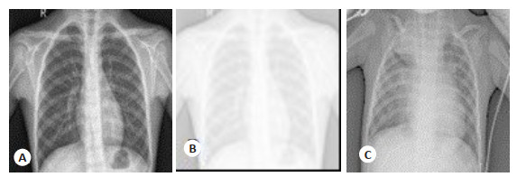
图 5 算法图像处理对比图
A:原始图像;B:SSR算法处理;C:改进算法处理
Figure 5. Algorithmic image processing comparison diagram.
表 1 Chest X-Ray Images数据集分布
Table 1. Distribution of chest X-ray images dataset
数据集 普通肺部图像 肺炎图像 合计 训练集 1341 3875 5216 测试集 234 390 624  下载: 导出CSV
表 2 Inception V3网络架构
Table 2. Network architecture of Inception V3
类型 Patch size/stride Or remark Input size Conv 3x3/2 299x299x3 Conv 3x3/1 149x149x32 Conv padded 3x3/1 147x147x32 Pool 3x3/2 147x147x64 Conv 3x3/1 73x73x64 Conv 3x3/2 71x71x80 Conv 3x3/1 35x35x192 3xInception 35x35x288 5xInception 7x17x768 2xInception 8x8x1280 Pool 8x8 8x8x2048 Linear Logits 1x1x2048 Softmax Classifier 1x1x1000
下载: 导出CSV
表 3 Inception V3网络第1次训练结果对比
Table 3. Comparison of the first training results of Inception V3 network
算法种类 准确性 特异性 敏感度 未使用 0.8766 0.7051 0.9795 SSR 0.8910 0.7436 0.9795 改进SSR 0.9779 0.7906 0.9846
下载: 导出CSV
表 4 Inception V3网络第2次训练结果对比
Table 4. Comparison of the second training results of Inception V3 network
算法种类 准确性 特异性 敏感度 未使用 0.8798 0.7308 0.9692 SSR 0.8798 0.7094 0.9861 改进SSR 0.9038 0.8077 0.9615
下载: 导出CSV
表 5 Inception V3网络第3次训练结果对比
Table 5. Comparison of the third training results of Inception V3 network
算法种类 准确性 特异性 敏感度 未使用 0.9038 0.8162 0.9564 SSR 0.8958 0.7949 0.9564 改进SSR 0.9215 0.8675 0.9538
下载: 导出CSV
表 6 Inception V3网络训练结果平均值
Table 6. Average value of network training results of perception V3
算法种类 准确性 特异性 敏感度 未使用 0.9038 0.8162 0.9564 SSR 0.8958 0.7949 0.9564 改进SSR 0.9215 0.8675 0.9538
下载: 导出CSV
-
[1] 唐丽. D-二聚体与肺炎病情评估的相关性[J]. 内蒙古医学杂志, 2017, 49(8): 929-31. https://www.cnki.com.cn/Article/CJFDTOTAL-NMYZ201708013.htm [2] 邓棋, 雷印杰, 田锋. 用于肺炎图像分类的优化卷积神经网络方法[J]. 计算机应用, 2020, 40(1): 71-6. doi: 10.3969/j.issn.1005-8451.2020.01.014 [3] Kermany DS, Goldbaum M, Cai W, et al. Identifying medical diagnoses and treatable diseases by image-based deep learning[J]. Cell, 2018, 172(5): 1122-31. e9. doi: 10.1016/j.cell.2018.02.010 [4] Vianna VP. Study and development of a computer-aided diagnosis system for classification of chest Xray images using convolutional neural networks pretrained for ImageNet and data augmentation [EB/OL]. [2018-09-16]. https://arxiv.org/pdf/1806.00839v1.pdf. [5] 何新宇, 张晓龙. 基于深度神经网络的肺炎图像识别模型[J]. 计算机应用, 2019, 39(6): 1680-4. https://www.cnki.com.cn/Article/CJFDTOTAL-JSJY201906022.htm [6] Kumar M, Gupta R, Raju KS, et al. Modified local binary pattern algorithm for feature dimensionality reduction[J]. Recent Adv Comput Sci Commun, 2021, 14(3): 934-40. doi: 10.2174/2213275912666190730160705 [7] 张亮, 王齐凯. 基于bp神经网络的肺炎分类[J]. 华南理工大学学报(自然科学版), 2015, 42(1): 72-6. doi: 10.3969/j.issn.1000-565X.2015.01.012 [8] Zhao G, Ahonen T, Matas J, et al. Rotation invariant image and video description with local binary pattern features[J]. IEEE T Image Process, 2011, 21(4): 1465-77 http://www.facweb.iitkgp.ernet.in/~debdoot/courses/EE60062/Aut2014/LBP.pdf [9] 朱珍, 万志平, 蒋鹏. 改进单尺度retinex的复杂光照人脸识别算法[J]. 计算机应用与软件, 2014, 31(3): 246-9, 262. doi: 10.3969/j.issn.1000-386x.2014.03.065 [10] Shrivastava J, Verma GS. Retinex Theory for Image Enhancement [J]. Res J Engi Tech, 2010, 1(2): 55-7. http://indianjournals.com/ijor.aspx?target=ijor:rjet&volume=1&issue=2&article=001 [11] 朱炼, 孙枫, 夏芳莉, 等. 图像融合研究综述[J]. 传感器与微系统, 2014, 33(2): 14-8. doi: 10.3969/j.issn.1000-9787.2014.02.004 [12] Perdomo O, Rios H, Rodríguez FJ, et al. Classification of diabetesrelated retinal diseases using a deep learning approach in optical coherence tomography[J]. Comput Methods Programs Biomed, 2019, 178: 181-9. doi: 10.1016/j.cmpb.2019.06.016 [13] Bhardwaj C, Jain S, Sood M. Diabetic retinopathy severity grading employing quadrant-based Inception-V3 convolution neural network architecture[J]. Int J Imaging Syst Technol, 2021, 31(2): 592-608. doi: 10.1002/ima.22510 [14] 汪泉, 宋文龙, 张怡卓, 等. 基于改进VGG16网络的机载高光谱针叶树种分类研究[J]. 森林工程, 2021, 37(3): 79-87. doi: 10.3969/j.issn.1006-8023.2021.03.011 [15] 刘长征, 相文波. 基于改进卷积神经网络的肺炎影像判别[J]. 计算机测量与控制, 2017, 25(4): 185-8. https://www.cnki.com.cn/Article/CJFDTOTAL-JZCK201704051.htm [16] Liu YF, Shu CY, Wang JD, et al. Structured knowledge distillation for dense prediction[J]. IEEE Trans Pattern Anal Mach Intell, 1940, PP(99): 1. http://arxiv.org/abs/1903.04197v5 [17] Shi T, Horvath S. Unsupervised learning with random forest predictors[J]. J Comput Graph Stat, 2006, 15(1): 118-38. doi: 10.1198/106186006X94072 [18] Zhang WP, Chen YR, Yang WM, et al. Class-variant margin normalized softmax loss for deep face recognition[J]. IEEE Trans Neural Netw Learning Syst, 2020: 1-6. http://www.researchgate.net/publication/343957338_Class-Variant_Margin_Normalized_Softmax_Loss_for_Deep_Face_Recognition [19] Khorami E, Mahdi BF, Azadeh A. Optimal Diagnosis of COVID-19 Based on Convolutional Neural Network and Red Fox Optimization Algorithm[J]. Comput Intell Neurosci, 2021: 4454507. DOI: 10.1155/2021/4454507. [20] Khorami E, Mahdi Babaei F, Azadeh A. Optimal diagnosis of COVID-19 based on convolutional neural network and red fox optimization algorithm[J]. Comput Intell Neurosci, 2021, 2021: 1- 11. [21] Ballesterp, Araujo RM. On the performance of GoogLeNet and AlexNet applied to sketches[C]// AAAI 2016: Proceedings of the 2016 Thirtieth AAAI Conference on Artificial Intelligence, Menlo Park, CA: AAAI, 2016: 1124-8. [22] 闫鹏程, 尚松行, 张超银, 等. 改进BP神经网络算法对煤矿水源的分类研究[J]. 光谱学与光谱分析, 2021, 41(7): 2288-93. https://www.cnki.com.cn/Article/CJFDTOTAL-GUAN202107056.htm [23] Parmar U, Pandya DH. Experimental investigation of cylindrical bearing fault diagnosis with SVM[J]. Mater Today: Proc, 2021, 44: 1286-90. doi: 10.1016/j.matpr.2020.11.327 [24] Perlin HA, Lopes HS. Extracting human attributes using a convolutional neural network approach[J]. Pattern Recognit Lett, 2015, 68: 250-9. doi: 10.1016/j.patrec.2015.07.012 [25] Zhang YJ. A Selection of Image Processing Techniques: From Fundamental to Research Front[M]. London. CRC Press: 2021. -

 点击查看大图
计量
- 文章访问数: 518
- HTML全文浏览量: 232
- PDF下载量: 42
- 被引次数: 0