


A new development in the application of resting functional magnetic resonance in temporal lobe epilepsy
-
摘要: 颞叶癫痫是成人最常见的局灶性癫痫。目前诊断癫痫的方法仍然以症状、电生理居多,脑电图检查中,发作期间的癫痫样放电是诊断癫痫的重要依据,但患者发作期较为短暂,捕捉异常脑电波信号具有一定困难,因此某种程度上,需要借助MRI进一步诊断。但仅依靠普通MRI检查在识别这些患者的癫痫发生区方面仍存在固有的困难。然而,借助改进的定位技术,颞叶癫痫通常可以正确识别。近年来,对于颞叶癫痫的研究已经转移到“功能”磁共振上。在“静止状态”下与大脑打交道。本文对目前可以分析颞叶癫痫患者静止状态的fMRI数据的诸多方法进行概述,包括低频振幅、基于种子的功能连接分析、区域同质性分析、独立成分分析、图分析等。Abstract: Temporal lobe epilepsy (TLE) is the most common focal epilepsy in adults. There are still inherent difficulties in identifying epileptic regions in these patients only by relying on ordinary MRI. However, with improved localization techniques, TLE can often be correctly identified. In recent years, the study of TLE has shifted to "functional" MRI. Working with the brain in a "static state". There are many ways to analyze fMRI data from the quiescent state of patients with TLE, and we will provide an extensive overview of them in this review.
-
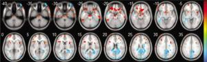
图 1 大脑的轴结构图
低频波动幅度的结果(静止状态功能磁共振成像); 暖色表示TLE>NC, 冷色表示NC>TLE.
Figure 1. Axial map of the brain.
-
[1] Chang YA, Marshall A, Bahrami N, et al. Differential sensitivity of structural, diffusion, and resting-state functional MRI for detecting brain alterations and verbal memory impairment in temporal lobe epilepsy[J]. Epilepsia, 2019, 60(5): 935-47. doi: 10.1111/epi.14736 [2] Haneef Z, Lenartowicz A, Yeh HJ, et al. Effect of lateralized temporal lobe epilepsy on the default mode network[J]. Epilepsy Behav, 2012, 25(3): 350-7. doi: 10.1016/j.yebeh.2012.07.019 [3] Liu M, Bernhardt BC, Hong SJ, et al. The superficial white matter in temporal lobe epilepsy: a key link between structural and functional network disruptions[J]. Brain, 2016, 139(pt 9): 2431-40. http://brain.oxfordjournals.org/content/139/9/2431 [4] Smitha KA, Akhil Raja K, Arun KM, et al. Resting state fMRI: a review on methods in resting state connectivity analysis and resting state networks[J]. Neuroradiol J, 2017, 30(4): 305-17. doi: 10.1177/1971400917697342 [5] Muhlhofer W, Tan YL, Mueller SG, et al. MRI-negative temporal lobe epilepsy-What do we know?[J]. Epilepsia, 2017, 58(5): 727-42. doi: 10.1111/epi.13699 [6] Korgaonkar MS, Ram K, Williams LM, et al. Establishing the resting state default mode network derived from functional magnetic resonance imaging tasks as an endophenotype: a twins study[J]. Hum Brain Mapp, 2014, 35(8): 3893-902. doi: 10.1002/hbm.22446 [7] Smith K. Brain imaging: fMRI 2.0[J]. Nature, 2012, 484(7392): 24-6. doi: 10.1038/484024a [8] Smith K. Brain decoding: Reading minds[J]. Nature, 2013, 502 (7472): 428-30. doi: 10.1038/502428a [9] Wang J, Shan Y, Dai J, et al. Altered coupling between resting-state glucose metabolism and functional activity in epilepsy[J]. Ann Clin Transl Neurol, 2020, 7(10): 1831-42. doi: 10.1002/acn3.51168 [10] Zhang ZQ, Lu GM, Zhong Y, et al. fMRI study of mesial temporal lobe epilepsy using amplitude of low-frequency fluctuation analysis [J]. Hum Brain Mapp, 2010, 31(12): 1851-61. doi: 10.1002/hbm.20982 [11] Singh TB, Aisikaer A, He C, et al. The assessment of brain functional changes in the temporal lobe epilepsy patient with cognitive impairment by resting-state functional magnetic resonance imaging [J]. J Clin Imaging Sci, 2020, 10: 50. doi: 10.25259/JCIS_55_2020 [12] Yang Z, Choupan J, Reutens D, et al. Lateralization of temporal lobe epilepsy based on resting-state functional magnetic resonance imaging and machine learning[J]. Front Neurol, 2015, 6: 184. [13] Bettus G, Bartolomei F, Confort-Gouny S, et al. Role of resting state functional connectivity MRI in presurgical investigation of mesial temporal lobe epilepsy[J]. J Neurol Neurosurg Psychiatry, 2010, 81 (10): 1147-54. doi: 10.1136/jnnp.2009.191460 [14] Sompairac N, Nazarov PV, Czerwinska U, et al. Independent component analysis for unraveling the complexity of cancer omics datasets[J]. Int J Mol Sci, 2019, 20(18): 4414. doi: 10.3390/ijms20184414 [15] Lv H, Wang Z, Tong E, et al. Resting-state functional MRI: everything that nonexperts have always wanted to know[J]. AJNR Am J Neuroradiol, 2018, 39(8): 1390-9. http://europepmc.org/abstract/MED/29348136 [16] Chassoux F, Artiges E, Semah F, et al. Determinants of brain metabolism changes in mesial temporal lobe epilepsy[J]. Epilepsia, 2016, 57(6): 907-19. doi: 10.1111/epi.13377 [17] Koch W, Teipel S, Mueller S, et al. Effects of aging on default mode network activity in resting state fMRI: Does the method of analysis matter?[J]. NeuroImage, 2010, 51(1): 280-7. doi: 10.1016/j.neuroimage.2009.12.008 [18] Michael AM, Anderson M, Miller RL, et al. Preserving subject variability in group fMRI analysis: performance evaluation of GICA vs. IVA[J]. Front Syst Neurosci, 2014, 26(8): 106. http://pubmedcentralcanada.ca/pmcc/articles/PMC4071815/ [19] Du Y, Allen EA, He H, et al. Artifact removal in the context of group ICA: a comparison of single-subject and group approaches[J]. Hum Brain Mapp, 2016, 37(3): 1005-25. doi: 10.1002/hbm.23086 [20] Du Y, Fan Y. Group information guided ICA for fMRI data analysis [J]. Neuroimage, 2013, 69: 157-97. doi: 10.1016/j.neuroimage.2012.11.008 [21] Erhardt EB, Rachakonda S, Bedrick EJ, et al. Comparison of multisubject ICA methods for analysis of fMRI data[J]. Hum Brain Mapp, 2011, 32(12): 2075-95. doi: 10.1002/hbm.21170 [22] Bartolomei F, Bettus G, Stam CJ, et al. Interictal network properties in mesial temporal lobe epilepsy: a graph theoretical study from intracerebral recordings[J]. Clin Neurophysiol, 2013, 124(12): 2345-53. doi: 10.1016/j.clinph.2013.06.003 [23] Stam CJ. Modern network science of neurological disorders[J]. Nat Rev Neurosci, 2014, 15(10): 683-95. doi: 10.1038/nrn3801 [24] Engel J, Thompson PM, Stern JM, et al. Connectomics and epilepsy [J]. Curr Opin Neurol, 2013, 26(2): 186-94. doi: 10.1097/WCO.0b013e32835ee5b8 [25] Constable RT, Scheinost D, Finn ES, et al. Potential use and challenges of functional connectivity mapping in intractable epilepsy [J]. Front Neurol, 2013, 4: 39. http://europepmc.org/articles/PMC3660665/ -

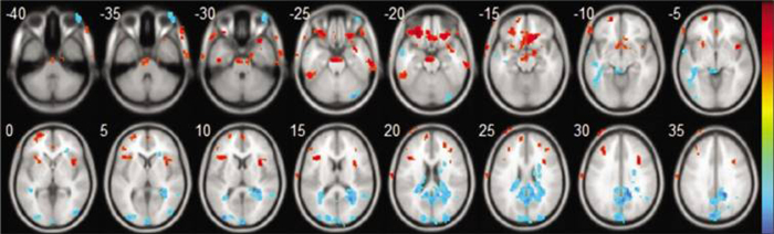
 下载:
下载:
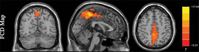
 点击查看大图
图(2)
计量
- 文章访问数: 444
- HTML全文浏览量: 179
- PDF下载量: 13
- 被引次数: 0