


Comparison of EUS, MRCP and CT in the diagnosis of stones < 4 mm in the lower segment of the bile duct
-
摘要:
目的对比超声内镜(EUS)与磁共振胰胆管造影(MRCP)、CT诊断胆系结石的临床价值。 方法回顾我院于2016年1月~2018年12月收治的165例可疑性胆系结石患者临床资料,其中男77例,女88例,年龄54~71岁(47.54±5.23岁),病程1~8年(4.24±0.58年)。分别采用EUS、MRCP、CT 3种检查方法对患者进行诊断,将诊断结果与手术病理诊断结果对比,分析3种诊断方法的敏感性、特异性、准确性、阳性预测值、阴性预测值以及不同直径结石的检出率。 结果在诊断胆系结石方面,EUS与MRCP的敏感性、特异性、准确性、阳性预测值、阴性预测值跟CT检查方式相比,差异有统计学意义(P < 0.05);EUS在胆系结石诊断方面的敏感性和准确性高于MRCP(P < 0.05);在 < 4 mm结石中,EUS检出率高于MRCP和CT,而MRCP检出率高于CT(P < 0.05)。 结论在胆系结石的诊断中,EUS、MRCP较CT效果更好,EUS较MRCP更具优势,尤其是对胆道下段 < 4 mm的微小结石。 Abstract:ObjectiveTo compare the clinical value of of EUS, MRCP and CT in the diagnosis of chololithiasis. MethodsEUS, MRCP and CT were used to diagnose 165 patients with suspected cholelithiasis. The diagnosis results were compared with the results of pathological diagnosis. The sensitivity, specificity, accuracy, PPV, NPV and detectionrate of stones with different diameterss of three diagnostic methods were analyzed and compared. ResultsIn the diagnosis of cholelithiasis, the sensitivity, specificity, accuracy, PPV and NPV of EUS and MRCP were significantly higher than CT (P < 0.05). The sensitivity and accuracy of EUS were significantly higher than MRCP (P < 0.05). For the stones < 4 mm, the detection rate of EUS was significantly higher than MRCP and CT (P < 0.05), while the detection rate of MRCP was significantly higher than CT (P < 0.05). ConclusionsIn the diagnosis of cholelithiasis, EUS and MRCP are significantly better than CT. EUS has more advantages over MRCP, especially for stones < 4 mm in the lower segment of the bile duct. -
Key words:
- cholelithiasis /
- endoscopic ultrasonography /
- magnetic resonance cholangiography /
- CT
-
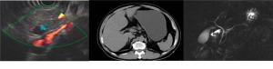
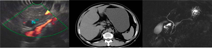
图 1 胆总管下段微小结石患者
A: EUS发现胆总管末端见一高回声, 确诊为胆总管下段微小结石; B: CT未发现结石; C: MRCP未发现结石
Figure 1. Atients with small stones in the lower part of the common bile duct
表 1 3种方法诊断结果分析(%)
Table 1. Analysis of diagnosis results of three methods
诊断方法 敏感性 特异性 准确性 阳性预测值 阴性预测值 EUS 97.5% (117/120)*# 95.75% (45/47)* 96.97% (160/165)*# 98.32% (117/119)* 95.56 (43/45)* MRCP 87.5% (105/120)* 91.84% (45/49)* 88.48% (146/165)* 96.33% (105/109)* 91.11 (41/45)* CT 73.33% (88/120) 80.34% (45/56) 73.33% (121/165) 88.89 (88/99) 75.56 (34/45) *P < 0.05 vs CT; #P < 0.05 vs MRCP.  下载: 导出CSV
表 2 3种方法不同直径结石检出率比较[n (%)]
Table 2. Comparison of stone detection rate in different diameter of three methods
结石直径 EUS MRCP CT ≥8 mm (n=24) 24 (100%) 23 (95.83%) 23 (95.83%) 4~7 mm (n=52) 51 (96.15%) 49 (92.31%) 40 (73.08%)* < 4 mm (n=44) 42 (93.19%) 33 (75%)# 25 (65.82%)*
下载: 导出CSV
-
[1] 宋睿, 杜国平.胆石症的研究进展与新认识[J].医学理论与实践, 2018, 31(10): 1427-8, 1436. http://d.old.wanfangdata.com.cn/Periodical/yxllysj201810009 [2] 朱军海. CT扫描和磁共振胰胆管成像在肝胆结石诊断中的应用价值比较[J].肝脏, 2016, 21(11): 1006-7. doi: 10.3969/j.issn.1008-1704.2016.11.033 [3] 刘雨, 胡良皞, 李兆申.胆管结石体外震波碎石研究进展[J].中华消化内镜杂志, 2019, 36(6): 450-3. doi: 10.3760/cma.j.issn.1007-5232.2019.06.016 [4] 赵文卓. MRI与CT在胆系结石疾病中的诊断分析[J].中国医药科学, 2019, 9(11): 140-2. doi: 10.3969/j.issn.2095-0616.2019.11.041 [5] 郑明伟.胆总管微小结石的影像学诊断[J].中国中西医结合外科杂志, 2017, 23(5): 572-5. doi: 10.3969/j.issn.1007-6948.2017.05.034 [6] 徐林生, 胡炳德, 梁丁保.超声内镜和磁共振胰胆管造影对胆总管结石诊断价值的对比研究[J].中华临床医师杂志:电子版, 2012, 6(21): 6876-7. http://d.old.wanfangdata.com.cn/Periodical/zhlcyszz201221070 [7] Lin LF, Huang PT. Linear endoscopic ultrasound for clinically suspected bile duct stones[J]. J Chin MedAssoc, 2012, 75(6): 251-4. [8] 卢青, 汪丽燕, 张剑波, 等. EUS和MRCP对胆总管结石的诊断价值[J].现代消化及介入诊疗, 2017, 22(5): 659-60. doi: 10.3969/j.issn.1672-2159.2017.05.014 [9] 张伟, 刘莉, 田英.胆总管结石应用CT、MRCP和超声内镜诊断的临床价值对比研究[J].中国CT和MRI杂志, 2019, 17(6): 89-91. doi: 10.3969/j.issn.1672-5131.2019.06.027 [10] 陈孝平.外科学[M].北京:人民卫生出版社, 2010. [11] 王超, 徐锋, 刘晓琳, 等.隐匿性胆总管结石诊断与治疗的新进展[J].临床肝胆病杂志, 2017, 33(7): 1391-6. doi: 10.3969/j.issn.1001-5256.2017.07.042 [12] del Pozo D, Tabernero S, Poves E, et al. Usefulness of endoscopic ultrasonography in the clinical suspicion of biliary disease[J]. Rev Esp Enferm Dig, 2011, 103(7): 345-8. doi: 10.4321/S1130-01082011000700002 [13] 李惠普. MRCP、超声内镜、CT在胆总管结石诊断中的应用价值[D].重庆: 重庆医科大学, 2017. http://cdmd.cnki.com.cn/Article/CDMD-10631-1017843811.htm [14] 高飞, 董江宁, 韦树华, 等. 3.0T MRI内插扰相快速梯度回波T1WI联合MRCP对胆系结石的诊断价值[J].实用放射学杂志, 2016, 32 (1): 60-3. doi: 10.3969/j.issn.1002-1671.2016.01.016 [15] Williams EJ, Green J, Beckingham I, et al. Guidelines on the management of common bile duct stones (CBDS)[J]. Gut, 2008, 57 (7): 1004-21. doi: 10.1136/gut.2007.121657 [16] Guarise A, Baltieri S, Mainardi P, et al. Diagnostic accuracy of MRCP in choledocholithiasis[J]. Radiol Med, 2005, 109(3): 239-51. http://d.old.wanfangdata.com.cn/Periodical/wcbxhgbxzz202002015 [17] Kondo S, Isayama H, Akahane M, et al. Detection of common bile duct stones: comparison between endoscopic ultrasonography, magnetic resonance cholangiography, and helical-computedtomographic cholangiography[J]. Eur J Radiol, 2005, 54(2): 271-5. doi: 10.1016/j.ejrad.2004.07.007 [18] 侯婧, 詹俊, 于钟, 等.两种胆胰管造影方法临床应用价值的荟萃分析[J].中华内科杂志, 2006, 45(11): 900-3. doi: 10.3760/j.issn:0578-1426.2006.11.007 [19] 金震东.超声内镜在消化系疾病诊治中的应用进展[J].胃肠病学和肝病学杂志, 2009, 18(1): 5-9. doi: 10.3969/j.issn.1006-5709.2009.01.002 [20] 姜宏雪, 郭杰芳, 金震东.超声内镜在胆系疾病诊治中的应用进展[J].中华消化内镜杂志, 2016, 33(8): 572-4. doi: 10.3760/cma.j.issn.1007-5232.2016.08.024 [21] 贾雷, 郭玉宁, 郭秀丽, 等.超声内镜与磁共振胰胆管造影诊断梗阻性黄疸的效果比较[J].临床肝胆病杂志, 2016, 32(9): 1753-5. doi: 10.3969/j.issn.1001-5256.2016.09.024 [22] 李惠普, 张俊文, 王谑菲. MRCP超声内镜CT在胆总管结石诊断中应用的meta分析[J].现代医药卫生, 2017, 33(5): 686-90. doi: 10.3969/j.issn.1009-5519.2017.05.014 [23] 张皞, 黄平, 张筱凤, 等.超声内镜、腹部超声及磁共振胰胆管造影对胆总管结石诊断价值的对比分析研究[J].中国内镜杂志, 2015, 21 (1): 26-9. http://d.old.wanfangdata.com.cn/Conference/8347921 [24] 陈流华, 郑朝旭, 谭敏, 等.内镜超声检查在腹腔镜胆囊切除术前的价值[J].中华腔镜外科杂志:电子版, 2010, 3(1): 102-7. http://d.old.wanfangdata.com.cn/Periodical/zhqjwkzz201001012 [25] 崔喻芳, 鲍峻峻, 宋育林, 等.超声内镜对胆总管结石的诊断价值[J].临床肝胆病杂志, 2019, 35(12): 2741-3. http://d.old.wanfangdata.com.cn/Periodical/lccsyxzz200506007 [26] 王楠, 石玉琪, 王帅, 等.胆总管结石患者临床诊断及治疗疗效分析[J].浙江临床医学, 2015, 32(8): 1333-4. http://d.old.wanfangdata.com.cn/Periodical/zjlcyx201508036 -

 点击查看大图
图(1) / 表(2)
计量
- 文章访问数: 797
- HTML全文浏览量: 469
- PDF下载量: 15
- 被引次数: 0