


Clinical application of 3D ASL in assessment of collateral circulation after unilateral internal carotid artery occlusion
-
摘要:
目的探讨三维动脉自旋标记(3D ASL)技术在评估单侧颈内动脉闭塞后侧支循环建立状态方面的临床应用价值。 方法收集2018年1月~2020年2月经三维时间飞跃法磁共振血管成像提示单侧颈内动脉闭塞且无其他颅内动脉中重度狭窄患者22例,其中男13例,女9例,年龄35~76岁(52.2±15.5岁),均行3D ASL序列灌注成像检查,使用Functool软件将原始数据自动生成脑血流量(CBF)伪彩图,分别于闭塞侧颈内动脉供血区及镜像区额叶、顶叶、脑室旁白质区、基底节区选取感兴趣区(ROI=200±20 mm2),并对比责任颈内动脉供血区与镜像区不同标记后延迟(PLD)时间脑血流量值差异。 结果入组患者中,左侧颈内动脉闭塞12例,右侧颈内动脉闭塞10例,3D ASL(PLD:1 525 ms)时闭塞颈内动脉供血区脑血流量值明显低于镜像区,两组差异有统计学意义(P<0.05),当PLD为2 525 ms时,闭塞颈内动脉供血区脑血流量值略低于镜像区,但差异无明显统计学意义(P>0.05)。 结论3D ASL成像技术可用于评估单侧颈内动脉闭塞后侧支循环建立及灌注状态,对于患者治疗方案的选择及预测临床预后均具有重要价值。 Abstract:ObjectiveTo evaluate the clinical value of 3D ASL in the assessment of collateral circulation after unilateral internal carotid artery (ICA) occlusion. MethodsFrom January 2018 to February 2020, 22 patients with unilateral ICA occlusion and no other moderate or severe stenosis of intracranial artery were studied by 3D-TOF MRA In ASL sequence perfusion imaging, including 13 males and 9 females with the age from 35 to 76 (average 52.2±15.5). The functool software was used to automatically generate the pseudocolor image of cerebral blood flow (CBF) from the original data. The areas of interest (ROI=200±20 mm2) were selected from the blood supply area of ICA on the occluded side and the frontal lobe, parietal lobe, paraventricular white matter area and basal ganglia area of the mirror image area. The CBF values of the time delay (PLD) between the responsible ICA blood supply area and the mirror image area were compared. ResultsThere were 12 cases of ICA occlusion on the left side and 10 cases of ICA occlusion on the right side. The CBF value in the blood supply area of ICA occlusion was significantly lower than that in the mirror area in 3D ASL (PLD: 1 525 ms). The difference between the two groups was significant (P<0.05). When PLD was 2 525 ms, the CBF value in the blood supply area of ICA occlusion was slightly lower than that in the mirror area, but the difference was not significant (P>0.05). Conclusion3D ASL imaging technology can be used to evaluate the establishment of collateral circulation and perfusion state after unilateral ICA occlusion, which is of great value for the selection of treatment plan and the prediction of clinical prognosis. -
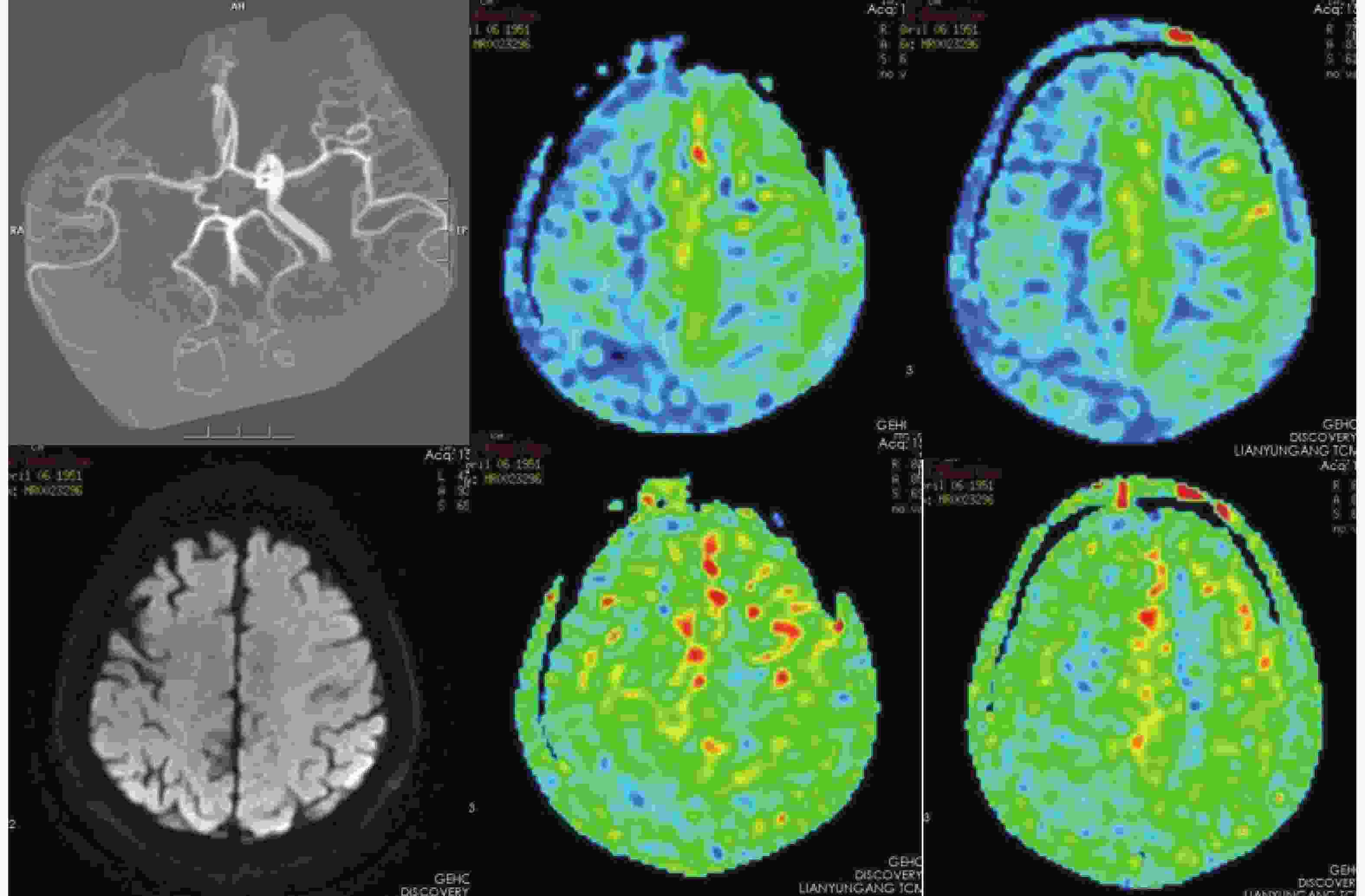
图 1 患者男,67岁,头晕1周,右侧颈内动脉闭塞后不同PLD CBF伪彩图
A: 3D-TOF MRA提示右侧ICA闭塞, ACA及右侧PCA开放, 右侧大脑后动脉偏侧优势; B-C: 3D ASL PLD:1 525 ms时提示右侧额顶叶低灌注; D: DWI序列未见异常信号; E-F: 3D ASL PLD:2 525 ms时提示双侧额顶叶脑血流量基本对称.
Figure 1. Patient, male ,67 years old, dizziness 1 week, different pseudo-color images after occlusion PLD CBF right internal carotid artery
表 1 不同PLD闭塞侧ICA供血区与镜像区CBF值 [mL/(min•100 g), Mean±SD]
Table 1. The CBF value of ICA blood supply area and mirror area in different occlusal side
部位 额叶CBF值 顶叶CBF值 侧脑室旁CBF值 基底节区CBF值 PLD:1 525 ms PLD:2 525 ms PLD:1 525 ms PLD:2 525 ms PLD:1 525 ms PLD:2 525 ms PLD:1 525 ms PLD:2 525 ms 责任供血区 22.562±8.316 50.462±5.847 21.265±7.836 49.554±4.763 19.741±6.972 40.841±5.225 19.863±7.254 40.872±5.652 镜像区 39.208±6.684 52.282±4.741 37.814±6.582 51.163±4.462 32.538±6.865 42.338±4.682 34.695±7.059 42.241±5.085 t −7.318 0 −1.134 0 −7.585 0 −1.156 3 −6.134 5 −1.000 8 −6.873 1 −0.844 6 P <0.001 0.263 2 <0.001 0.254 1 <0.001 0.322 6 <0.001 0.403 1 CBF: 脑血流量;ICA: 颈内动脉; PLD: 标记后延迟.  下载: 导出CSV
-
[1] 赵仁军, 刘 英, 杨加惠. TCD检测颈内动脉颅外段重度狭窄或闭塞后颅内血流动力学改变的临床价值[J]. 中西医结合心脑血管病杂志, 2017, 15(13): 1653-5. doi: 10.3969/j.issn.1672-1349.2017.13.037 [2] 常佩佩, 苗延巍, 蒋玉涵, 等. 单侧大脑中动脉狭窄患者FLAIR血管高信号与三维动脉自旋标记动脉内穿行伪影的一致性及其影响因素[J]. 中国医学影像技术, 2019, 35(10): 1456-60. [3] 李 晨, 何 文, 杜丽娟, 等. 经颅CEUS评价单侧颈内动脉颅外段重度狭窄或闭塞患者脑灌注成像的可行性[J]. 中国介入影像与治疗学, 2016, 13(4): 234-9. [4] 范晓媛, 冯 逢. 动脉自旋标记MRI技术在烟雾病中的应用[J]. 国际医学放射学杂志, 2019, 42(6): 668-72. [5] 郑园园, 惠品晶, 韩佳霖, 等. 经颅多普勒量化评估单侧颈内动脉重度狭窄或闭塞侧支循环的可行性[J]. 中风与神经疾病杂志, 2018, 35(9): 782-6. [6] 李义, 张保朝, 曹伟光, 等. DSA对单侧颈内动脉系统大动脉狭窄或闭塞后侧支循环建立的应用价值[J]. 中国实用神经疾病杂志, 2017, 20(5): 33-6. doi: 10.3969/j.issn.1673-5110.2017.05.011 [7] 雷少阳, 张淑倩. ASL技术在中枢神经系统中的应用进展[J]. 国际医学放射学杂志, 2020, 43(1): 73-7. [8] Kaczmarz S, Griese V, Preibish C, et al. Increased variability of watershed areas in patients with high-grade carotid stenosis[J]. Neuroradiology, 2018, 60(3): 313-3. [9] 刘 洋, 王拓一, 何冬若, 等. 颈总动脉闭塞性病变的血流代偿模式及意义[J]. 中华老年心脑血管病杂志, 2019, 21(12): 1256-9. doi: 10.3969/j.issn.1009-0126.2019.12.007 [10] 陆文杰, 肖正光, 顾 峰, 等. 基于MR定量积分及血液动力学参数评价颈动脉粥样硬化同脑梗死的相关性[J]. 医学影像学杂志, 2018, 28(6): 903-7. [11] 张萍淑, 吴小英, 钱琳琳, 等. 慢性单侧颈内动脉闭塞患者脑动脉血流动力学变化观察[J]. 中国综合临床, 2019, 35(3): 199-203. doi: 10.3760/cma.j.issn.1008-6315.2019.03.002 [12] 张萍淑, 吴小英, 孔祥慧, 等. 慢性单侧颈内动脉颅外段闭塞患者脑动脉血流分流观察[J]. 山东医药, 2017, 57(43): 68-70. doi: 10.3969/j.issn.1002-266X.2017.43.021 [13] Liu CH, Chang CH, Chang TY, et al. Carotid artery stenting improves cerebral hemodynamics regardless of the flow direction of ophthalmic artery[J]. Angiology, 2015, 66(2): 180-6. doi: 10.1177/0003319714522854 [14] 林天烨, 有 慧, 冯 逢, 等. 动脉自旋标记MR技术进展及应用[J]. 中华放射学杂志, 2019, 53(5): 431-4. [15] 游润发, 周海军. 动脉自选标记技术在常见脑部疾病中的应用进展[J]. 中华神经医学杂志, 2019, 18(1): 93-7. doi: 10.3760/cma.j.issn.1671-8925.2019.01.018 [16] Ma H, Wang Z, Xu K, et al. Three-dimensional arterial spin labeling imaging and dynamic susceptibility contrast perfusion-weighted imaging value in diagnosing glioma grade prior to surgery[J]. Exp Ther Med, 2017, 13(6): 2691-8. doi: 10.3892/etm.2017.4370 [17] 赵 辉, 闫俊强, 张育德, 等. MRI动脉自旋标记技术在脑血管疾病诊断中的应用[J]. 中国CT和MRI杂志, 2018, 16(8): 15-7. doi: 10.3969/j.issn.1672-5131.2018.08.005 [18] 吴秀美, 严江志, 王健, 等. 颈内动脉狭窄患者动脉自旋标记成像临床应用研究进展[J]. 中华老年心脑血管病杂志, 2019, 21(10): 1117-20. doi: 10.3969/j.issn.1009-0126.2019.10.030 [19] 周倩, 王倩倩, 刘新疆. 磁共振三维动脉自旋标记技术研究进展及临床应用[J]. 磁共振成像, 2019, 10(12): 955-60. [20] Zhang J, Xia C, Liu Y, et al. Comparative study of MR mTI-ASL and DSC -PWI in evaluating cerebral hemodynamics of patients with Moyamoya disease[J]. Med Baltim, 2018, 97(41): 12768-75. doi: 10.1097/MD.0000000000012768 [21] 刘征华, 刘征春, 祝莎莎. 基于多期ASL成像技术的单侧大脑中动脉狭窄的ATT等研究[J]. 临床放射学杂志, 2019, 38(9): 1594-8. [22] Lou X, Yu S, Scalzo F, et al. Multi-delay ASL can identify leptomeningeal collateral perfusion in endovascular therapy of ischemic stroke[J]. Oncotarget, 2017, 8(2): 2437-43. [23] 周建国, 符大勇, 马先军, 等. ASL对大脑中动脉M1段闭塞后侧支循环建立显示的临床应用[J]. 实用放射学杂志, 2018, 34(8): 1164-6, 1171. doi: 10.3969/j.issn.1002-1671.2018.08.003 [24] Haga S, Morioka T, Shimogawa T, et al. Arterial spin labeling perfusion magnetic resonance image with dual postlabeling delay : a correlative study with acetazolamide loading (123) I-Iodoamphetamine single-photon emission computed tomography[J]. Stroke Cerebrovasc Dis, 2016, 25(1): 1-6. doi: 10.1016/j.jstrokecerebrovasdis.2015.08.025 [25] 刘松国, 韩 广, 于秀英, 等. 不同后标记延迟三维动脉自旋标记技术对大脑中动脉狭窄的评估[J]. 中国中西医结合影像学杂志, 2019, 17(3): 231-7. doi: 10.3969/j.issn.1672-0512.2019.03.004 -

 点击查看大图

图(1) / 表(1)
计量
- 文章访问数: 736
- HTML全文浏览量: 352
- PDF下载量: 10
- 被引次数: 0