


Preliminary application of artificial intelligence-assisted CT in screening and monitoring of COVID-19
-
摘要:
目的探讨人工智能辅助CT在COVID-19病变筛查以及病情监测评估中的应用价值。 方法收集27例COVID-19患者的CT影像资料,其中男性14例,女性13例,年龄28~85岁(48.9±14.3岁)。将图像输入基于深度学习模型的“uAI新冠肺炎智能辅助分析系统”,软件自动批量进行肺炎病灶识别和标记,并自动计算病变总体积、内部磨玻璃影体积及实变区域体积。通过PACS系统对人工智能辅助诊断软件识别病灶进行人工诊断复核,记录软件识别区域假阳性或假阴性情况,并通过手动修复少数假阳性或假阴性图像。 结果人工智能辅助诊断软件可对肺炎病灶进行自动识别和标记,并计算出患者病灶总体积、内部磨玻璃影体积及实变区域体积。通过人工复核诊断显示人工智能辅助诊断软件对病灶标记的范围与肉眼观察相比具有较好的一致性。20例临床普通型患者均未见假阳性或假阴性病例;重症及危重症患者中有3例患者可见局部软件标记病灶呈假阳性表现,临床患者类型组间的差异有统计学意义(P<0.05)。人工智能辅助诊断软件提供的随访功能可直观的以图片及图表方式呈现两次检查病灶范围及密度变化的对比情况。人工复核诊断显示2例患者可见局部病灶标识区域呈假阴性表现,3例患者可见假阳性表现,临床患者类型组间的差异有统计学意义(P<0.05)。 结论人工智能辅助CT可有效识别COVID-19病灶,并提供病灶相关数据信息。在患者病情评估方面通过图片及图表方式可直观的显示病变范围及内部密度差异的变化,为临床评效提供客观数据支持,同时提高了影像医师的工作效率。 Abstract:ObjectiveTo explore the application value of artificial intelligence-assisted CT in the screening of the novel coronavirus pneumonia and the monitoring. MethodsCT imaging data of 27 patients with novel coronavirus pneumonia were collected, including 14 males and 13 females with the age from 28 to 85 years old (average 48.9±14.3). The imagings were loaded to the "uAI novel coronavirus pneumonia Intelligent Assisted Analysis System" based on deep learning models. Then the software automatically identified and labeled the pneumonia lesions in batches, and automatically calculated the total volume of lesions, the volume of internal ground glass shadow and the volume of consolidation area. After that, the PACS system was used to manually review the diagnosis of lesions identified by the artificially assisted diagnosis software, record the false positive or false negative situation in the software recognition area, and manually repair a few false positive or false negative images. ResultsArtificial intelligence-assisted diagnosis software automatically identified and labeled the pneumonia lesions. It calculated the total volume of the patient's lesions, the volume of the internal ground glass shadow and the volume of the consolidation area. The results of manual reexamination showed that the range of lesions labeled by the artificial intelligence -assisted diagnosis software was more consistent with that observed by the naked eye. There were no false-positive or false-negative cases in 20 clinical general-type patients. Among the severe and critical patients, 3 patients showed false positive manifestations of local software-labeled lesions, and the difference between patients in different clinical types was significant (P<0.05). The follow-up function provided by the artificial intelligence-assisted diagnosis software visually showed the comparison of the changes in the range and density of the two lesions by the forms of pictures and charts. The manual review diagnosis showed that 2 patients presented false negative manifestations in the local lesion labeled area, and 3 patients presented false positive manifestations, and the difference between patients in different clinical types was significant (P<0.05). ConclusionArtificial intelligence-assisted CT can effectively identify the lesions of novel coronavirus pneumonia and provide detail information about the lesions. In terms of the assessment of patients' condition, the changes in the lesion range and internal density differences can be visually shown by pictures and charts, which provide objective data support for clinical evaluation and improve the work efficiency of imaging physicians. -
Key words:
- COVID-19 /
- artificial intelligence /
- CT /
- screening /
- condition monitoring
-
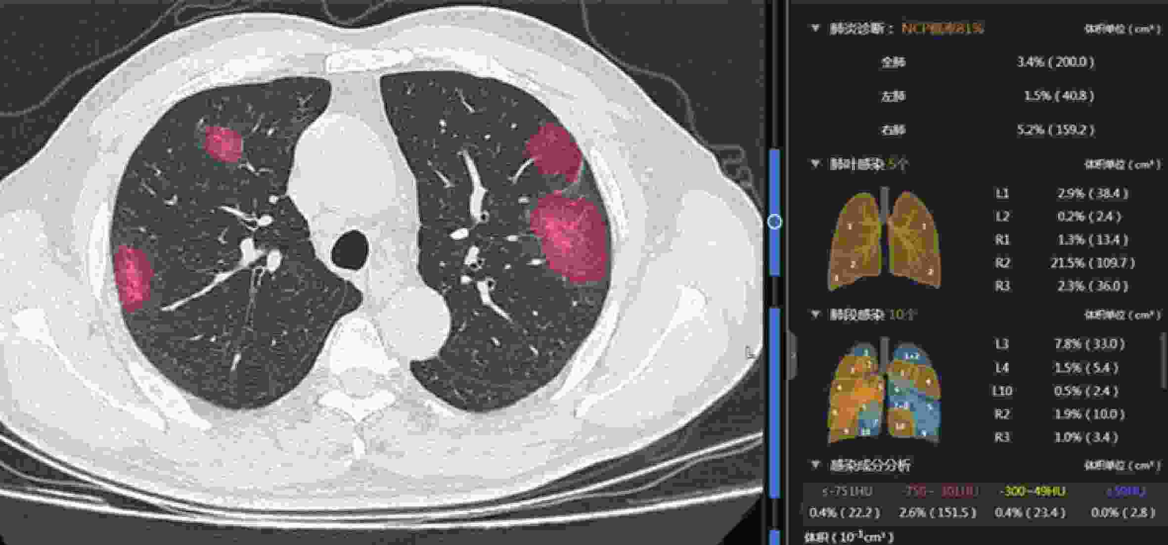
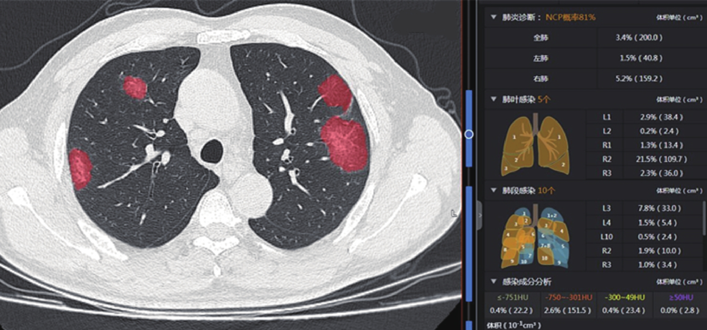
图 1 人工智能辅助诊断软件示意图
患者男,58岁,确诊COVID-19,人工智能辅助诊断软件对肺炎病灶进行自动识别、标记,并计算出病灶总体积及相关密度体积.
Figure 1. Diagrammatic sketch of artificial intelligence auxiliary diagnostic software
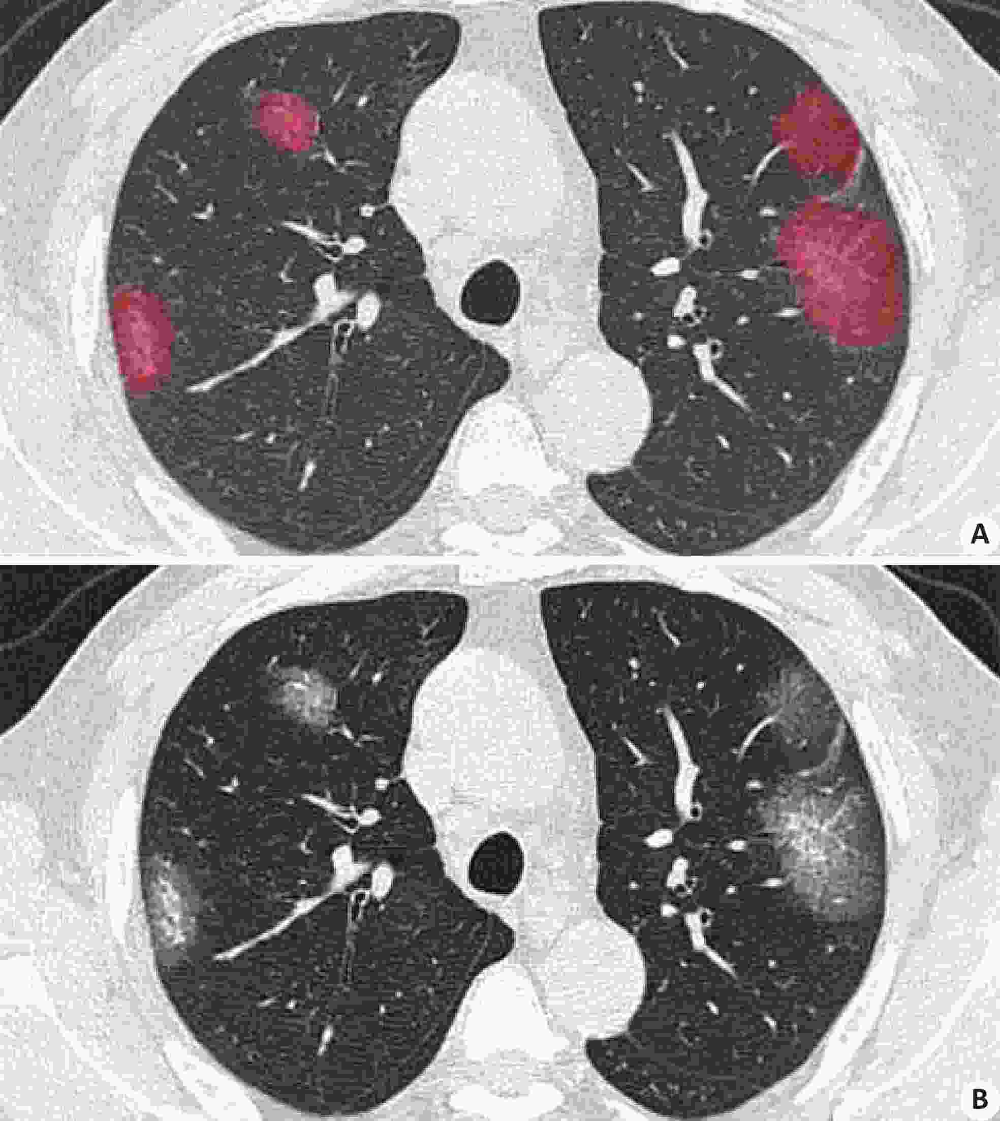
图 2 人工智能辅助诊断软件病灶标记范围与人工复核肉眼观察对比
患者男,58岁,确诊COVID-19,人工智能辅助诊断软件病灶标记范围与人工复核肉眼观察相比一致性较高.
Figure 2. Comparison of the range of focal markings of artificial intelligence assisted diagnostic software and artificial review of naked eye observation
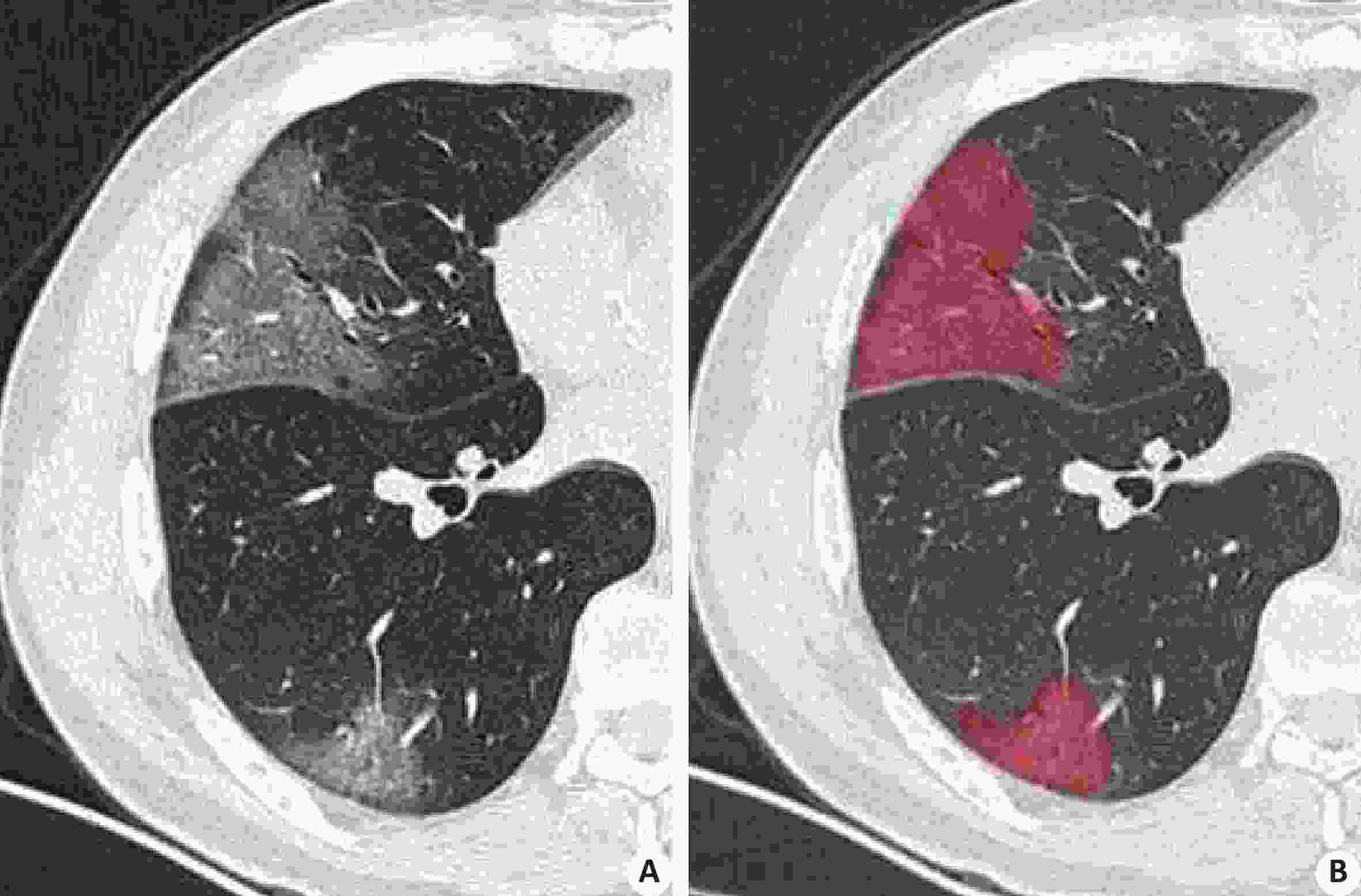
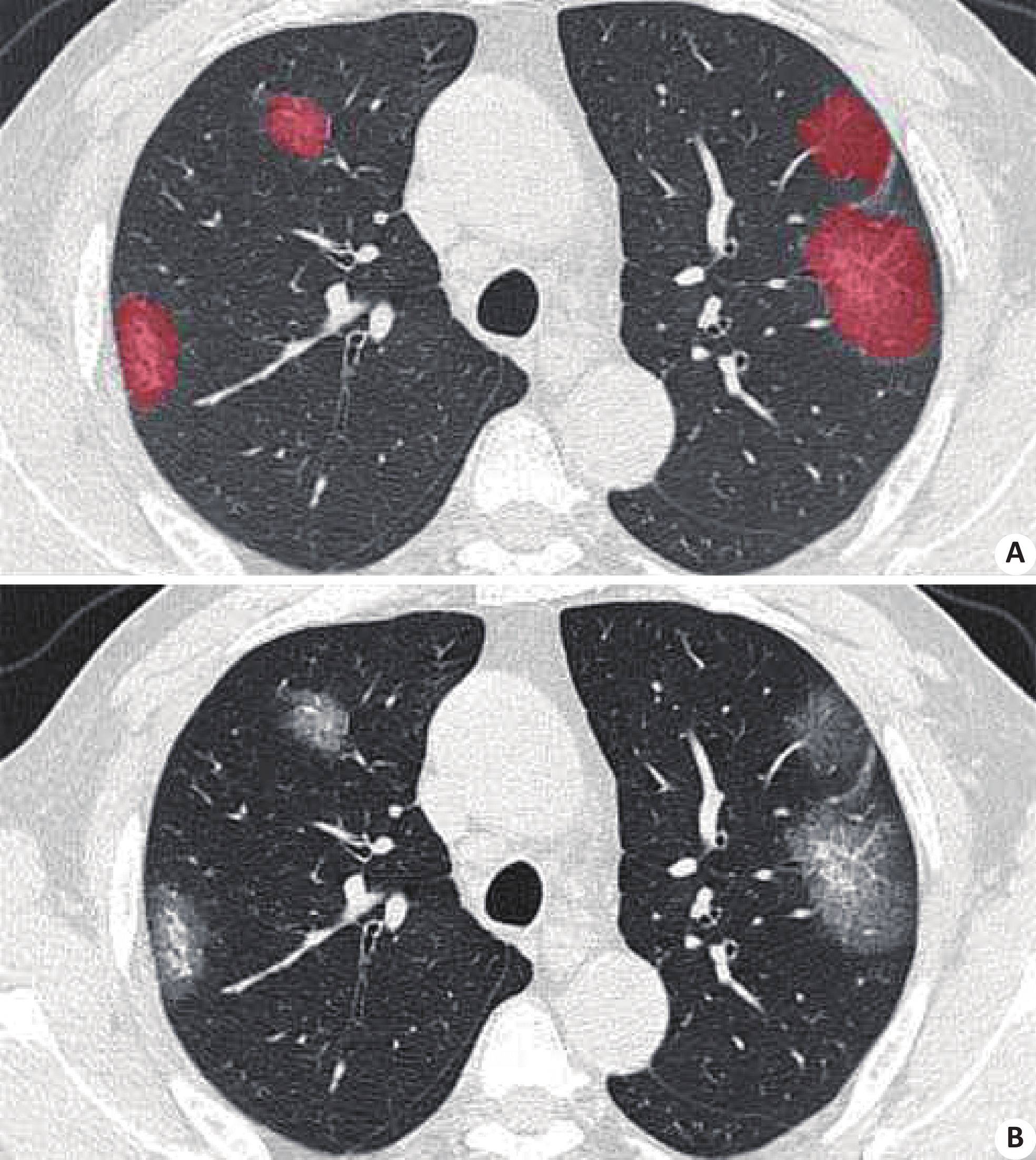
图 3 人工智能辅助诊断软件病灶标记范围与人工复核肉眼观察对比
患者男,49岁,确诊COVID-19,人工智能辅助诊断软件病灶标记范围与人工复核肉眼观察相比一致性较高.
Figure 3. Comparison of the range of focal markings between artificial intelligence assisted diagnostic software and artificial review of naked eye observation
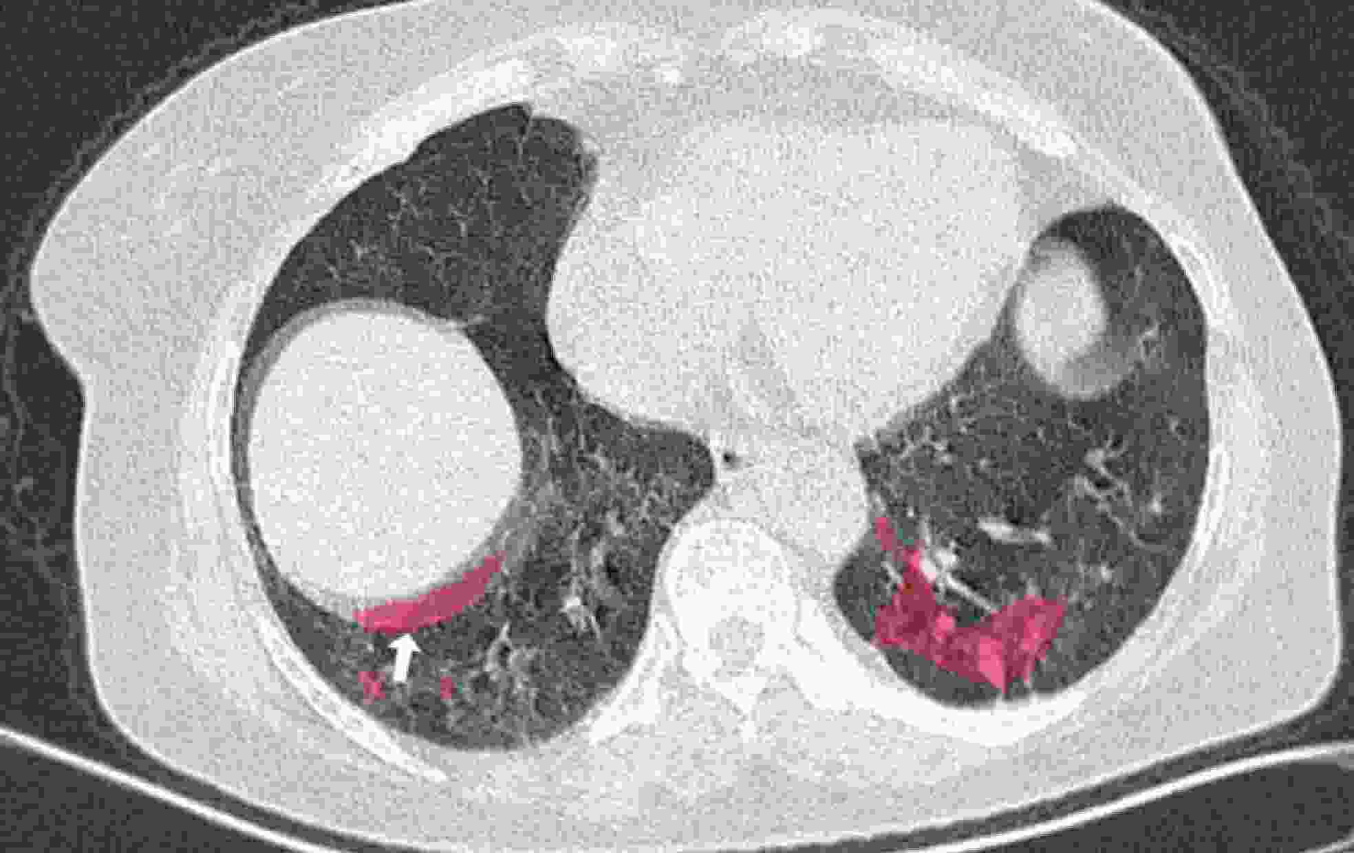
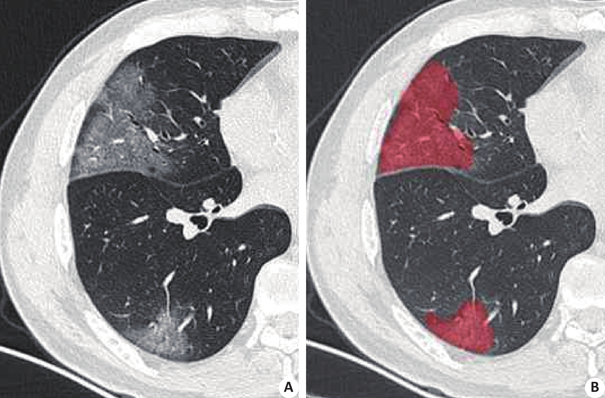
图 4 人工智能辅助诊断软件标注病灶局部呈假阳性表现
患者女,62岁,确诊COVID-19。患者扫描时屏气不佳导致图像出现容积伪影,被人工智能辅助诊断软件误标为病灶.
Figure 4. Local false positive features of lesions marked by artificial intelligence assisted diagnostic software
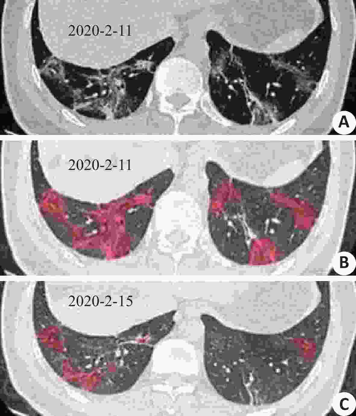
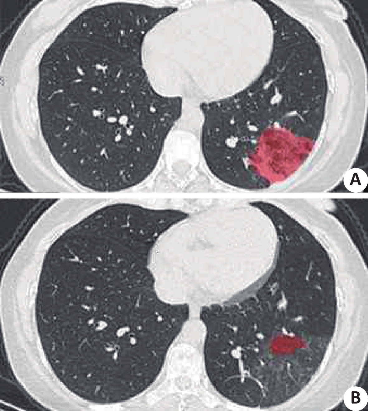
图 5 人工智能辅助诊断软件示意图
患者男,42岁,确诊COVID-19,CT复查显示病灶范围缩小,局部见残留纤维条索,人工智能辅助诊断软件显示病灶标记区范围缩小.
Figure 5. Diagrammatic sketch of the artificial intelligence auxiliary diagnostic software
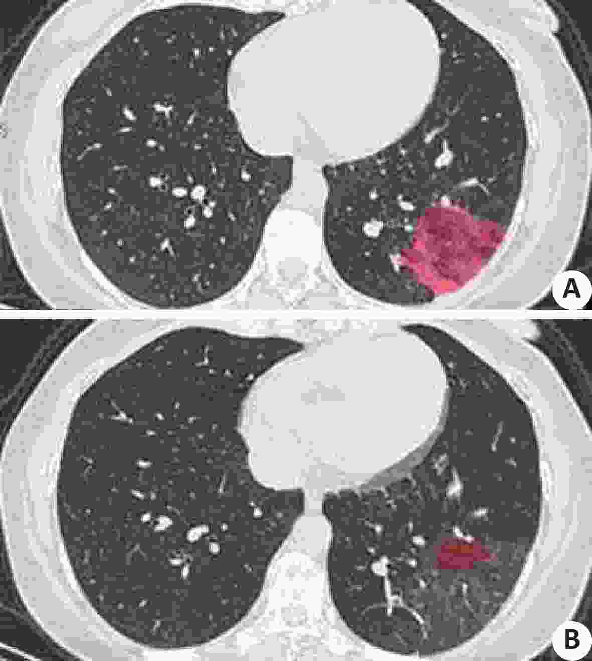
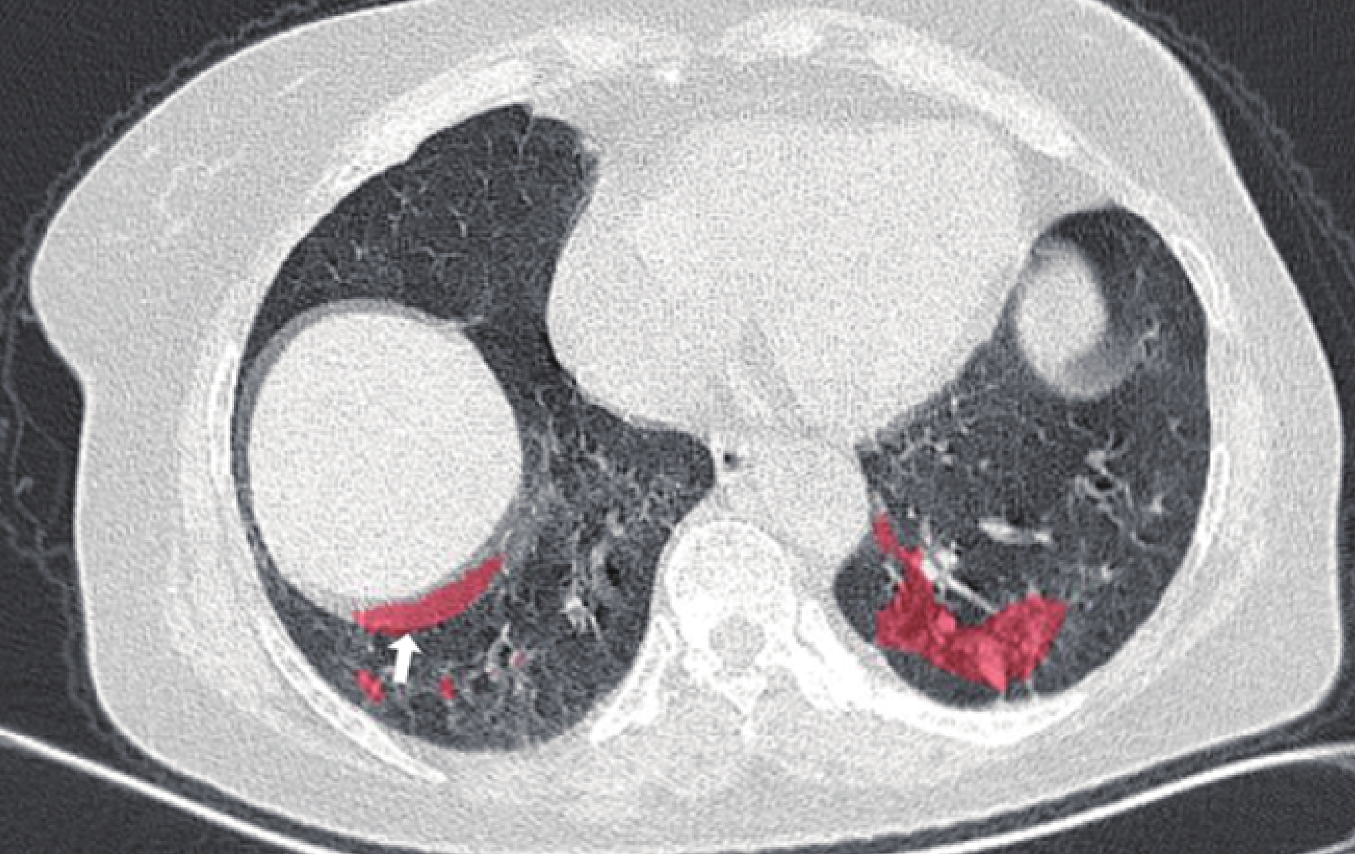
图 6 人工智能辅助诊断软件标记病灶呈假阴性表现
患者女,29岁,确诊新COVID-19,原病灶区复查肉眼观察见轻微片絮状稍高密度影,对应区域软件未予以标记.
Figure 6. The artificial intelligence auxiliary diagnostic software marked false negative manifestations of focal points
表 1 COVID-19患者相关数据分析
Table 1. Data analysis COVID-19 patients
序号 性别 年龄(岁) 临床分型 累及肺叶数目 首次CT检查 CT复查 CT复查影像评估 病变体积(cm3) 磨玻璃(cm3) 实性(cm3) 病变体积(cm3) 磨玻璃(cm3) 实性(cm3) 1 男 41 普通型 4 132.1 68.3 47.9 11.3 6.9 2.9 好转 2 女 33 普通型 3 214.1 109.2 74.8 117.7 60.5 34.6 好转 3 男 63 重型 5 822.3 382.1 257.8 948.4 568.3 275.6 进展 4 男 58 普通型 5 200.1 151.5 23.4 196.2 147.4 31.6 无明显变化 5 男 52 普通型 5 248.9 115.5 92.9 168.3 51.8 79.8 好转 6 男 62 危重型 5 1 185.3 633.9 186.8 1393.1 798.7 362.6 进展 7 女 45 普通型 4 276.6 57.7 138.3 261.1 62.1 133.9 无明显变化 8 女 67 重型 5 409.8 267.6 76.6 465.1 239.1 141.5 进展 9 女 28 普通型 2 77.3 37.3 23.3 10.7 6.9 0.7 好转 10 男 45 普通型 2 17.6 10.2 3.1 4.9 3.0 0.4 好转 11 女 49 普通型 2 36.2 9.8 17.5 6.0 2.8 2.1 好转 12 男 66 普通型 5 235.1 124.6 66.6 50.9 33.6 11.3 好转 13 女 51 危重型 5 952.3 663.8 184.2 1436.5 529.6 629.1 进展 14 男 31 重型 5 1 225.6 734.3 287.4 851.5 557.6 132.2 好转 15 女 46 普通型 5 308.1 169.4 72.9 152.6 86.9 34.0 好转 16 男 35 普通型 4 46.1 30.8 8.9 20.7 12.1 3.8 好转 17 男 29 普通型 1 1.7 0.9 0.6 0.3 0.3 0 好转 18 女 30 普通型 2 21.2 13.5 6.5 1.3 1.0 0.2 好转 19 女 85 重型 5 771.6 436.8 232.3 1233.2 637.2 417.6 进展 20 女 49 普通型 4 28.6 19.2 4.9 16.2 11.5 2.6 好转 21 男 67 普通型 3 151.7 87.2 45.3 41.8 10.3 20.9 好转 22 女 37 普通型 2 7.7 4.5 1.2 4.0 2.8 0.3 好转 23 男 41 普通型 4 27.6 20.3 3.3 18.8 14.1 1.9 好转 24 女 54 普通型 5 76.5 40.4 22.7 7.3 2.3 2.5 好转 25 男 49 普通型 5 286.6 159.8 98.6 201.4 128.3 44.8 好转 26 女 65 普通型 3 121.4 57.1 54.4 91.5 50.1 26.5 好转 27 男 42 重型 5 539.2 309.3 102.8 367.1 170.0 113.4 好转  下载: 导出CSV
表 2 COVID-19智能辅助分析软件病灶标识与人工诊断复核符合情况(n)
Table 2. Conformance of COVID-19 intelligent assisted analysis software focus identification and artificial diagnosis review
临床分型 首次CT 复查CT 符合 不符合 符合 不符合 普通型 20 0 19 1 重型及危重型 4 3 3 4 χ2 9.643 9.343 P 0.002 0.002
下载: 导出CSV
-
[1] Pan F, Ye TH, Sun P, et al. Time course of lung changes on chest CT during recovery from 2019 novel coronavirus (COVID-19) pneumonia[J]. Radiology, 2020. https://pubs.rsna.org/doi/10.1148/radiol.2020200370. [2] Zu ZY, Jiang MD, Xu PP, et al. Coronavirus disease 2019(COVID-19): a perspective from China[J]. Radiology, 2020. https://pubmed.ncbi.nlm.nih.gov/32083985/. [3] Pan YY, Guan HX, Zhou SC, et al. Initial CT findings and temporal changes in patients with the novel coronavirus pneumonia (2019-nCoV): a study of 63 patients in Wuhan, China[J]. Eur Radiol, 2020. DOI: 10.1007/s00330-020-06731-x. [4] 管汉雄, 熊 颖, 申楠茜, 等. 新型冠状病毒肺炎(COVID-19)临床影像学特征[J]. 放射学实践, 2020, 35(2): 125-30. [5] 李欣菱, 郭芳芳, 周 振, 等. 基于深度学习的人工智能胸部CT肺结节检测效能评估[J]. 中国肺癌杂志, 2019, 22(6): 336-40. doi: 10.3779/j.issn.1009-3419.2019.06.02 [6] 明佳蕾, 方向明. 基于人工智能的CT肺结节检出临床应用及研究进展[J]. 中华放射学杂志, 2019, 53(6): 522-5. [7] Sun WQ, Zheng B, Qian W. Automatic feature learning using multichannel ROI based on deep structured algorithms for computerized lung cancer diagnosis[J]. Comput Biol Med, 2017, 89: 530-9. doi: 10.1016/j.compbiomed.2017.04.006 [8] 王 浩, 萧 毅, 孟祥峰, 等. 医学影像人工智能临床使用质量控制[J]. 中华放射学杂志, 2019, 53(9): 723-7. doi: 10.3760/cma.j.issn.1005-1201.2019.09.002 [9] 李小虎, 潘红利, 束晶苇, 等. 输入性新型冠状病毒肺炎临床和CT特征[J]. 中国医学影像技术, 2020, 36(2): 248-51. [10] Chung M, Bernheim A, Mei XY, et al. CT imaging features of 2019 novel coronavirus (2019-nCoV)[J]. Radiology, 2020, 295(1): 202-7. doi: 10.1148/radiol.2020200230 [11] Lei JQ, Li JF, Li X, et al. CT imaging of the 2019 novel coronavirus (2019-nCoV) pneumonia[J]. Radiology, 2020, 295(1): 18. doi: 10.1148/radiol.2020200236 [12] Fang YC, Zhang HQ, Xu YY, et al. CT manifestations of two cases of 2019 novel coronavirus (2019-nCoV) pneumonia[J]. Radiology, 2020, 295(1): 208-9. doi: 10.1148/radiol.2020200280 [13] Shi HS, Han XY, Zheng CS. Evolution of CT manifestations in a patient recovered from 2019 novel coronavirus (2019-nCoV) pneumonia in Wuhan, China[J]. Radiology, 2020, 295(1): 20. doi: 10.1148/radiol.2020200269 [14] 许 强, 张其锐, 卢光明. 新一代医学影像人工智能临床转化现状与挑战[J]. 中华放射学杂志, 2019, 53(11): 913-5. [15] 蔡雅倩, 张正华, 韩 丹, 等. AI对肺磨玻璃结节筛查及定性的临床应用研究[J]. 放射学实践, 2019, 34(9): 958-62. [16] 王成弟, 郭际香, 杨 阳, 等. 利用深度学习技术辅助肺结节的人工智能检测[J]. 中国呼吸与危重监护杂志, 2019, 18(3): 288-94. [17] 张惠茅, 萧 毅, 洪 楠, 等. 医学影像人工智能产业现状和发展需求调研报告[J]. 中华放射学杂志, 2019, 53(6): 507-11. [18] 刘士远, 萧 毅. 基于深度学习的人工智能对医学影像学的挑战和机遇[J]. 中华放射学杂志, 2017, 51(12): 899-901. doi: 10.3760/cma.j.issn.1005-1201.2017.12.002 -


 点击查看大图
计量
- 文章访问数: 857
- HTML全文浏览量: 350
- PDF下载量: 13
- 被引次数: 0