


Evaluation of ventricular function changes before and after anthracycline treatment in patients with breast cancer by three-dimensional speckle tracking echocardiography
-
摘要:
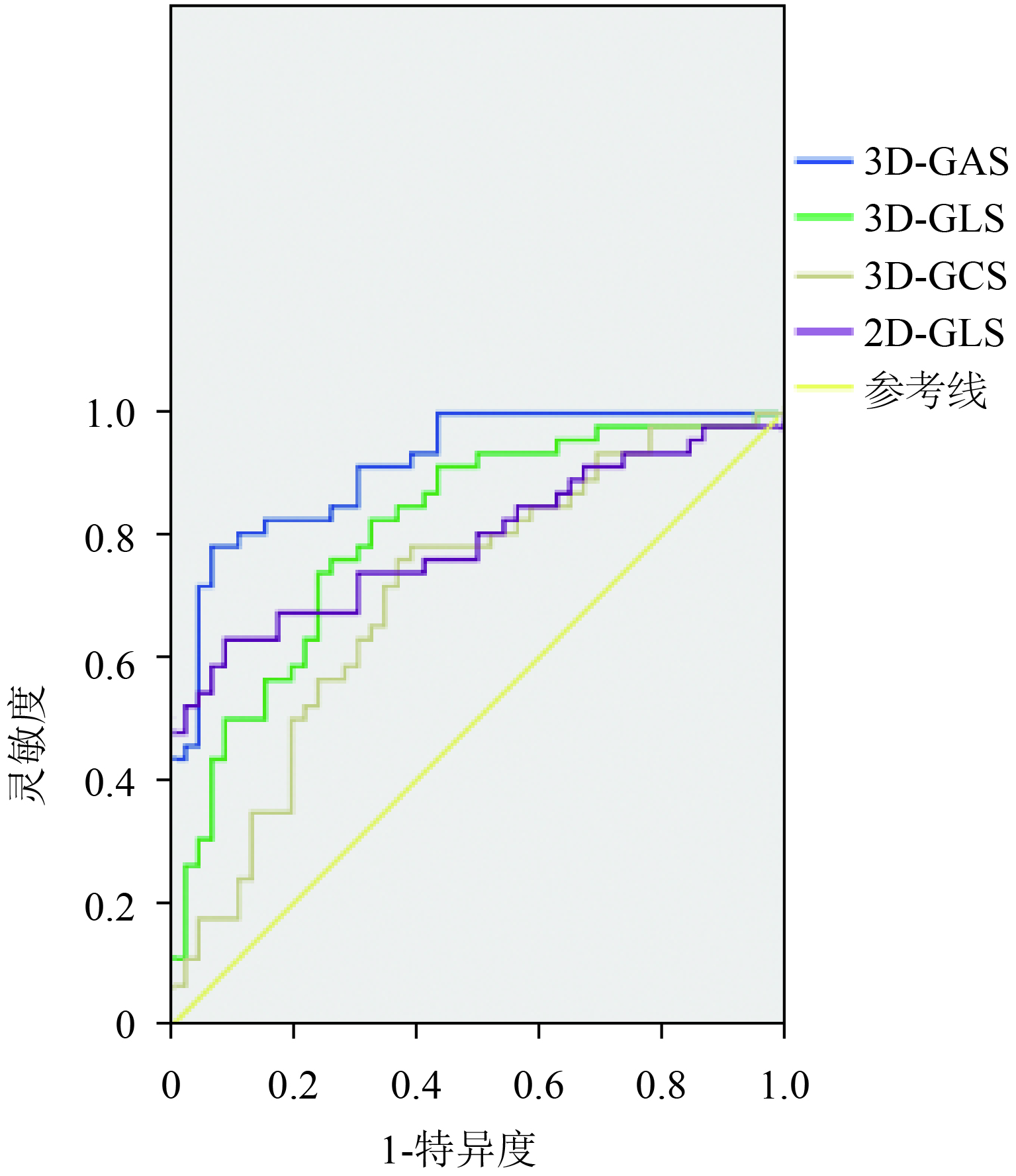
目的探讨三维斑点追踪技术(3D-STE)评价乳腺癌患者蒽环类药物治疗前后心室功能改变。 方法应用常规超声心动图、二维斑点追踪技术(2D-STE)、3D-STE分别在蒽环类药物化疗前24 h、化疗第2周期及化疗第4周期结束24 h内进行左心室功能检测,对各参数在这3个时间点进行纵向对比,分析其与蒽环类药物累积剂量的相关性,以及对检测蒽环类药物心脏毒性所致心脏功能早期损害的敏感性。 结果3D-STE的左室整体面积应变(3D-GAS)、整体长轴应变(3D-GLS)在化疗第2周期与化疗第4周期结束24 h内均低于化疗前24 h(P<0.05);左室整体圆周应变(3D-GCS)在化疗第4周期结束24 h内低于化疗前24 h(P<0.05);而左室整体径向应变(3D-GRS)于整个化疗过程中无明显改变(P>0.05);在检测左心室功能早期损害方面,3D-GAS的敏感性为81.23%,特异性为81.98%;3D-GLS的敏感性为62.15%,特异性为65.02%;3D-GCS的敏感性为66.09%,特异性为55.08%;3D-GAS、3D-GLS、3D-GCS与蒽环类药物累积剂量成负相关(P<0.05)。 结论3D-STE评价乳腺癌患者蒽环类药物治疗前后心室功能改变值得在临床影像学诊断中推广使用,是监测蒽环类药物所致心脏功能损害的一种敏感、可靠的影像学手段。 Abstract:ObjectiveTo explore three-dimensional speckle tracking echocardiography (3D-STE) in evaluating the ventricular function changes in breast cancer patients before and after the administration of anthracyclines. MethodsRoutine echocardiography, two-dimensional speckle tracking echocardiography (2D-STE) and 3D-STE were separately applied to test the left ventricular function 24 h before, within 24 h of the end of the 2nd and the 4th cycle of anthracycline chemotherapy. The parameters obtained at these 3 time points were compared longitudinally. The correlation with the cumulative dose of anthracyclines was analyzed. Their sensitivity to the detection of early heart impairment caused by cardiotoxicity of anthracycline was analyzed. ResultsThe left ventricular global area strain (3D-GAS) and the global longitudinal strain (3D-GLS) within 24 h after the end of the 2nd and 4th cycle of chemotherapy were lower than those before chemotherapy (P<0.05). The left ventricular global circumferential strain (3D-GCS) within 24 h after the end of the 4th cycle of chemotherapy was lower than that before 24 h of chemotherapy (P<0.05). The change in global radial strain (3D-GRS) during the whole chemotherapy process was not significant (P>0.05). The sensitivity and specificity for 3D-GAS were 81.23% and 81.98%, respectively; 62.15% and 65.02% for 3D-GLS, respectively; 66.09% and 55.08% for 3D-GCS, respectively. 3D-GAS, 3D-GLS, 3D-GCS were negatively correlated with the cumulative dose of anthracyclines(P<0.05). Conclusion3D-STE for evaluating ventricular function changes in breast cancer patients before and after using anthracyclines is worth popularizing in clinical imaging diagnosis. It is a sensitive and reliable imaging method for monitoring the heart impairment caused by anthracyclines. -
Key words:
- three-dimensional spot tracking /
- breast cancer /
- anthracyclines /
- ventricular function
-
表 1 化疗前24 h、化疗第2周期及化疗第4周期结束24 h内常规超声心动图参数变化比较(Mean±SD)
化疗时期 IVSd(mm) LVDd(mm) LVPWd(mm) LVEF(%) E/A 化疗前24 h 8.83±0.69 46.04±1.97 8.39±0.72 64.09±3.98 1.22±0.19 化疗第2周期结束24 h内 8.38±0.67 45.32±1.55 8.32±0.89 62.24±2.89 1.16±0.23 化疗第4周期结束24 h内 8.98±0.91 46.23±1.93 8.46±0.63 61.74±2.26 1.19±0.28 IVSd:室间隔舒张末期厚度;LVDd:左室舒张末期内径;LVPWd:左室后壁舒张末期厚度;LVEF:左室射血分数  下载: 导出CSV
表 2 化疗前24 h、化疗第2周期及化疗第4周期结束24 h内3D-STE与2D-STE各应变参数比较(Mean±SD)
化疗时期 3D-GAS 3D-GLS 3D-GCS 3D-GRS 2D-GLS 化疗前24 h −34.19±2.47 −18.43±2.48 −18.45±2.67 −34.18±4.54 −20.34±2.07 化疗第2周期结束24 h内 −29.13±2.09* −16.39±1.90* −17.29±3.79 −33.21±4.93 −20.19±1.67 化疗第4周期结束24 h内 −24.89±2.41* −15.29±1.91* −15.79±3.65* −32.06±5.09 −18.88±1.25* *P<0.05 vs化疗前24 h
下载: 导出CSV
-
[1] Carvalho FS, Burgeiro A, Garcia R, et al. Doxorubicin-Induced cardiotoxicity: from bioenergetic failure and cell death to cardiomyopathy[J]. Med Res Rev, 2014, 34(1): 106-35. doi: 10.1002/med.21280 [2] 胡夕春, 张 剑, 陈德滇, 等. 中国蒽环类药物治疗乳腺癌专家共识[J]. 中国肿瘤临床, 2018, 55(3): 120-5. doi: 10.3969/j.issn.1000-8179.2018.03.198 [3] Florescu M, Cinteza M, Vinereanu D. Chemotherapy-induced cardiotoxicity[J]. Maedica (Buchar), 2013, 8(1): 59-67. [4] Mladosievicova B, Urbanova D, Radvanska E, et al. Role of NT-proBNP in detection of myocardial damage in childhood leukemia survivors treated with and without anthracyclines[J]. J Exp Clin Cancer Res, 2012, 31(2): 86-97. [5] Carver JR, Szalda D, Ky B. Asymptomatic cardiac toxicity in Long-Term cancer survivors: defining the population and recommendations for surveillance[J]. Semin Oncol, 2013, 40(2): 229-38. doi: 10.1053/j.seminoncol.2013.01.005 [6] Conrad AL, Gundrum JD, Mchugh VL, et al. Utility of routine left ventricular ejection fraction measurement before anthracycline-based chemotherapy in patients with diffuse large B-cell lymphoma[J]. J Oncol Pract, 2012, 8(6): 336-40. doi: 10.1200/JOP.2012.000682 [7] Chung WB, Yi JE, Jin JY, et al. Early cardiac function monitoring for detection of subclinical Doxorubicin cardiotoxicity in young adult patients with breast cancer[J]. J Breast Cancer, 2013, 16(2): 178-83. doi: 10.4048/jbc.2013.16.2.178 [8] Baysal T, Koksal Y, Oran B, et al. Cardiac functions evaluated with tissue Doppler imaging in childhood cancers treated with anthracyclines[J]. Pediatr Hematol Oncol, 2010, 27(1): 13-23. doi: 10.3109/08880010903352299 [9] Rajapreyar P, Lorenzana A, Prabhu A, et al. Tissue doppler imaging and focal, late-onset anthracycline-induced cardiovascular disease in long term survivors of childhood cancer: a research article[J]. J Clin Diagn Res, 2016, 10(8): C1-4. [10] 郑言言, 王 玲, 陈剑琼, 等. 斑点追踪技术评价化疗对癌症患者左心功能的影响[J]. 中国超声医学杂志, 2012, 28(11): 999-1002. doi: 10.3969/j.issn.1002-0101.2012.11.016 [11] Stoodley PW, Richards DA, Boyd A, et al. Altered left ventricular longitudinal diastolic function correlates with reduced systolic function immediately after anthracycline chemotherapy[J]. Eur Heart J Cardiovasc Imaging, 2013, 14(3): 228-34. doi: 10.1093/ehjci/jes139 [12] Nesser HJ, Mor-Avi V, Gorissen W, et al. Quantification of left ventricular volumes using three-dimensional echocardiographic term left ventricular remodelling after acute myocardial infarction[J]. Eur J Nucl Med Mol Imaging, 2011, 38(6): 1124-31. doi: 10.1007/s00259-011-1739-7 [13] Senju N, Ikeda S, Koga S, et al. The echocardiographic Tei-index reflects early myocardial damage induced by anthracyclines in patients with hematological malignancies[J]. Heart Vessels, 2007, 22(6): 393-7. doi: 10.1007/s00380-007-0985-x [14] Shaikh AY, Suryadevara S, Tripathi AA, et al. Mitoxantrone-Induced cardiotoxicity in acute myeloid LeukemiaA velocity vector imaging analysis[J]. Echocardiography, 2016, 33(8): 1166-77. doi: 10.1111/echo.13245 [15] Rohde LE, Baldi A, Weber C, et al. Tei index in adult patients submitted to adriamycin chemotherapy: failure to predict early systolic dysfunction. Diagnosis of adriamycin cardiotoxicity[J]. Int J Cardiovasc Imaging, 2007, 23(2): 185-91. doi: 10.1007/s10554-006-9145-0 [16] Zhu PH, Huang JY, Ye M, et al. Assessment of left ventricular twist in type 2 diabetes mellitus by using two-dimensional ultrasound speckle tracking imaging[J]. J Zhejiang Univ(Med Sci), 2014, 43(5): 566-71. [17] Woo JS, Yu TK, Kim WS, et al. Early prediction of myocardial viability after acute myocardial infarction by two-dimensional speckle tracking imaging[J]. J Geriatr Cardiol, 2015, 12(5): 474-81. [18] de Almeida AL, Silva VA, de Souza FA, et al. Subclinical ventricular dysfunction detected by speckle tracking two years after use of anthracycline[J]. Arq Bras Cardiol, 2015, 104(4): 274-83. [19] Guerra F, Marchesini M, Contadini D, et al. Speckle-tracking global longitudinal strain as an early predictor of cardiotoxicity in breast carcinoma[J]. Support Care Cancer, 2016, 24(7): 3139-45. [20] Wang W, Kang Y, Shu XH, et al. Early detection of the cardiotoxicity induced by chemotherapy drug through two-dimensional speckle tracking echocardiography combined with high-sensitive cardiac troponin T[J]. Chin J Oncol, 2017, 39(11): 835-40. [21] Tang Q, Jiang Y, Xu Y, et al. Speckle tracking echocardiography predicts early subclinical anthracycline cardiotoxicity in patients with breast cancer[J]. J Clin Ultrasound, 2017, 45(4): 222-30. doi: 10.1002/jcu.22434 [22] 张小花, 姜志荣, 李大海, 等. 蒽环类药物对肿瘤患者左心功能影响的组织多普勒评价[J]. 临床超声医学杂志, 2007, 9(3): 151-3. doi: 10.3969/j.issn.1008-6978.2007.03.008 [23] 礼广森, 任卫东, 崔洪岩, 等. 定量组织速度成像评价乳腺癌患者表阿霉素心脏毒性左心室功能的变化[J]. 中国超声医学杂志, 2006, 22(12): 908-10. doi: 10.3969/j.issn.1002-0101.2006.12.010 [24] Lotrionte M, Cavarretta E, Abbate A, et al. Temporal changes in standard and tissue Doppler imaging echocardiographic parameters after anthracycline chemotherapy in women with breast cancer[J]. Am J Cardiol, 2013, 112(7): 1005-12. doi: 10.1016/j.amjcard.2013.05.038 -

 点击查看大图
图(2) / 表(2)
计量
- 文章访问数: 687
- HTML全文浏览量: 352
- PDF下载量: 12
- 被引次数: 0