


Diffuse large B-cell lymphoma of the esophagus and cardia complicated with bronchogenic carcinoma: a case report and literature review
-
摘要:
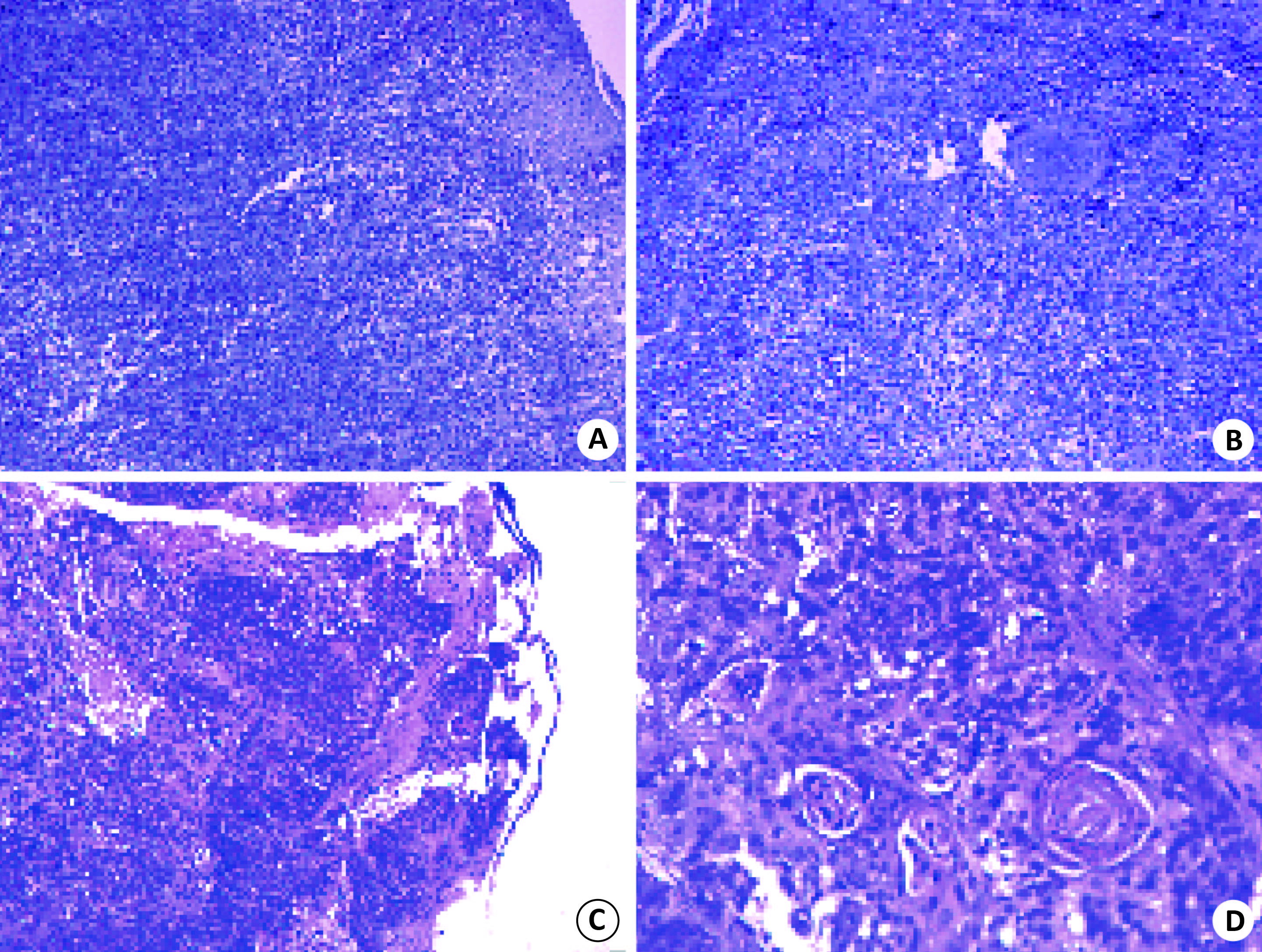
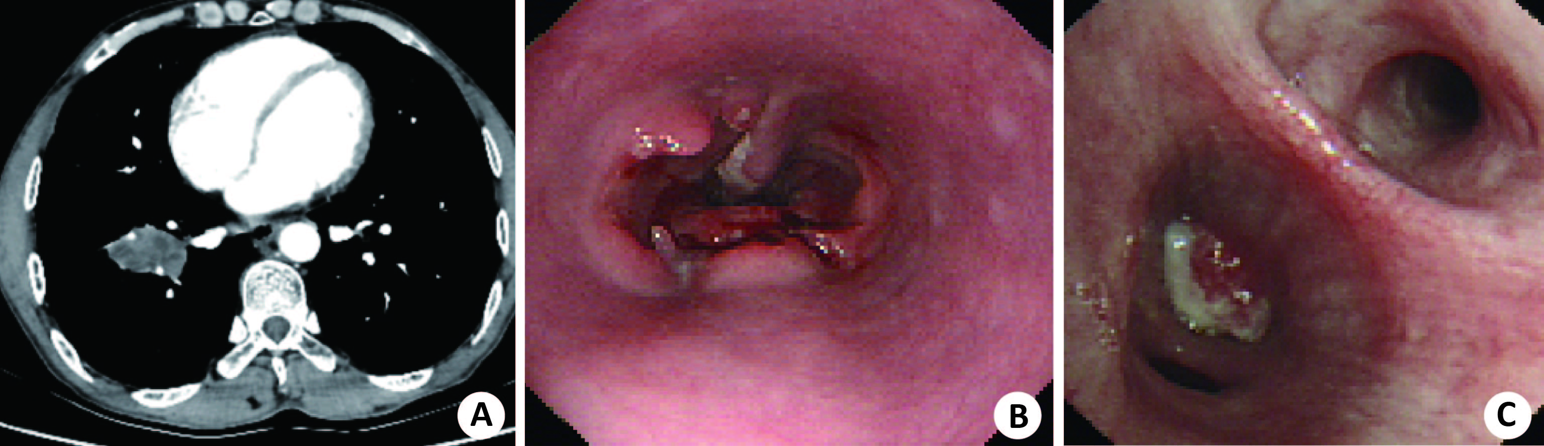
目的 探讨食管贲门弥漫性大B细胞淋巴瘤合并原发性支气管肺癌的诊治过程,分析其容易误诊的原因,为临床提供借鉴。 方法回顾性分析1例食管贲门弥漫性大B细胞淋巴瘤合并原发性支气管肺癌患者的病例资料及诊治过程,并结合相关文献进行分析。 结果患者入院后完善胸腹部增强CT、胃镜检查,考虑为贲门恶性肿瘤侵及食管、肺继发恶性肿瘤,遂行支气管镜检查,活检结果提示贲门食管部弥漫性大B细胞淋巴瘤合并原发性支气管肺癌。 结论食管弥漫性大B细胞淋巴瘤合并支气管肺癌时极易形成食管气管瘘,严重危及患者生命,故发现食管溃疡性病变合并肺部占位性病变时应行食管及肺部病理活检,以明确诊断,制定下一步治疗方案。 -
关键词:
- 食管 /
- 贲门 /
- 弥漫性大B细胞淋巴瘤 /
- 非霍奇金淋巴瘤 /
- 支气管肺癌
Abstract:Objective To investigate the diagnosis and treatment of diffuse large B-cell lymphoma of esophagus and cardia complicated with primary bronchogenic carcinoma, and analyze the causes of misdiagnosis, provide reference for clinical practice. Methods The clinical data, diagnosis and treatment of a patient with diffuse large B-cell lymphoma of esophagus and cardia complicated with primary bronchogenic carcinoma were analyzed retrospectively. The related literatures were analyzed. Results After admission, enhanced CT and gastroscopy were performed in the chest and abdomen. The bronchoscopy was performed for the secondary malignant tumor of esophagus and lung involved by cardiac malignant tumor. The biopsy results suggested that diffuse large B-cell lymphoma of the cardia and esophagus was associated with primary bronchogenic carcinoma. Conclusion Diffuse large B-cell lymphoma complicated with bronchogenic carcinoma is easy to form esophagotracheal fistula, which seriously endangers the life of the patients. Therefore, pathological biopsy of esophagus and lung should be performed when esophageal ulcerative lesions complicated with pulmonary space-occupying lesions found. It can help make a definite diagnosis and make the next treatment plan. -
Key words:
- esophagus /
- cardia /
- diffuse large B cell lymphoma /
- non hodgkin lymphoma /
- bronchogenic carcinoma
-
[1] Li S, Young KH, Medeiros LJ. Diffuse large B-cell lymphoma[J]. Pathology, 2018, 50(1): 74-87. doi: 10.1016/j.pathol.2017.09.006 [2] Lee YY, Takata K, Wang RC, et al. Primary gastrointestinal anaplastic large cell lymphoma[J]. Pathology, 2017, 49(5): 479-85. doi: 10.1016/j.pathol.2017.05.007 [3] Armitage JO. Staging non-Hodgkin lymphoma[J]. CA Cancer J Clin, 2005, 55(6): 368-76. doi: 10.3322/canjclin.55.6.368 [4] 张 用, 毕建平, 皮国良, 等. 国际肺癌研究协会第八版国际肺癌TNM分期修订稿解读[J]. 肿瘤防治研究, 2016, 43(4): 313-8. doi: 10.3971/j.issn.1000-8578.2016.04.016 [5] 支修益, 石远凯, 于金明. 中国原发性肺癌诊疗规范(2015年版)[J]. 中华肿瘤杂志, 2015, 37(1): 67-78. doi: 10.3760/cma.j.issn.0253-3766.2015.01.014 [6] 张宏娜. 胃肠道淋巴瘤临床研究进展[J]. 中国微创外科杂志, 2018, 18(5): 450-3. doi: 10.3969/j.issn.1009-6604.2018.05.017 [7] Manning MA, Somwaru AS, Mehrotra AK, et al. Gastrointestinal lymphoma: radiologic-pathologic correlation[J]. Radiol Clin North Am, 2016, 54(4): 765-78. doi: 10.1016/j.rcl.2016.03.007 [8] Gurney KA, Cartwright RA. Increasing incidence and descriptive epidemiology of extranodal non-Hodgkin lymphoma in parts of England and Wales[J]. Hematol J, 2002, 3(2): 95-104. doi: 10.1038/sj.thj.6200154 [9] Jirapat, Teerakanok, Ju dy, et al. Primary esophageal diffuse large B cell lymphoma presenting with tracheoesophageal fistula: a rare case and review[J]. World J Gastrointest Oncol, 2017, 9(10): 431-5. doi: 10.4251/wjgo.v9.i10.431 [10] Prasanna, Ghimire. Primary esophageal lymphoma in immunocompetent patients: two case reports and literature review[J]. World J Radiol, 2010, 2(8): 334-8. doi: 10.4329/wjr.v2.i8.334 [11] Berman MD, Falchuk KR, Trey C, et al. Primary histiocytic lymphoma of the esophagus[J]. Dig Dis Sci, 1979, 24(11): 883-6. doi: 10.1007/BF01324907 [12] Tirumani H, Rosenthal MH, Tirumani SH, et al. Imaging of uncommon esophageal malignancies[J]. Dis Esophagus, 2015, 28(6): 552-9. doi: 10.1111/dote.12208 [13] Ma Q, Zhang C, Fang S, et al. Primary esophageal mucosa-associated lymphoid tissue lymphoma: a case report and review of literature[J]. Medicine(Baltimore), 2017, 96(13): e6478-86. [14] Chadha KS, Hernandez-Ilizaliturri FJ, Javle M. Primary esophageal lymphoma: case series and review of the literature[J]. Dig Dis Sci, 2006, 51(1): 77-83. doi: 10.1007/s10620-006-3088-0 [15] Fujishima F, Katsushima H, Fukuhara NA, et al. Incidence rate, subtype frequency, and occurrence site of malignant lymphoma in the gastrointestinal tract: population-based analysis in miyagi, Japan[J]. Tohoku J Exp Med, 2018, 245(3): 159-65. doi: 10.1620/tjem.245.159 [16] Yu L, Chen K, Xu Y, et al. The value of EUS in combination with cytological, flow cytometry, and gene rearrangement in the diagnosis of gastrointestinal lymphoma[J]. Hematol Oncol, 2017, 35(3): 303-9. doi: 10.1002/hon.v35.3 [17] Rohatiner A, Damore F, Coiffier B, et al. Report on a workshop convened to discuss the pathological and staging classifications of gastrointestinal tract lymphoma[J]. Ann Oncol, 1994, 5(5): 397-400. doi: 10.1093/oxfordjournals.annonc.a058869 [18] 孔智渊. 胃底贲门腺癌合并回盲部弥漫大B细胞淋巴瘤1例[J]. 医药前沿, 2013, 34(19): 339-40. doi: 10.3969/j.issn.2095-1752.2013.19.405 [19] Thieblemont C, Zucca E. Clinical aspects and therapy of gastrointestinal MALT lymphoma[J]. Best Pract Res Clin Haematol, 2017, 30(1/2): 109-17. [20] Nakamura S, Matsumoto T. Gastrointestinal lymphomas: recent topics in diagnosis and treatment[J]. Nihon Shokakiby Gakk Zassh, 2017, 114(11): 1933-8. [21] Floch P, Megraud F, Lehours P. Helicobacter pylori strains and gastric MALT lymphoma[J]. Toxins(Basel), 2017, 9(4): 513-23. [22] Espinoza JL, Matsumoto A, Tanaka HA. Gastric microbiota: an emerging player in helicobacter pylori-induced gastric malignancies[J]. Cancer Lett, 2018, 414(1): 147-52. [23] Naoki, Asano, Katsunori, et al. Helicobacter pylori-negative gastric mucosa-associated lymphoid tissue lymphomas: a review[J]. World J Gastroenterol, 2015, 21(26): 8014-20. doi: 10.3748/wjg.v21.i26.8014 [24] Xia Y, Zhou Z. Treatment of primary gastric lymphoma[J]. Chin J Gastrointest Surg, 2017, 20(11): 1227-30. [25] Aviles A, Castaneda C, Cleto S, et al. Rituximab and chemotherapy in primary gastric lymphoma[J]. Cancer Biother Radiopharm, 2009, 24(1): 25-7. doi: 10.1089/cbr.2008.0507 [26] 刘祥祥, 王健红, 郝彩霞, 等. 17例原发胃肠道弥漫大B细胞淋巴瘤临床病例分析[J]. 现代肿瘤医学, 2018, 26(17): 2756-9. doi: 10.3969/j.issn.1672-4992.2018.17.028 [27] 仲恺励, 张伟京. 原发胃恶性淋巴瘤诊治进展[C]//第八届全国难治性白血病学术研讨会, 2012: 13-8. [28] 郑 建, 高 红, 洪成雨, 等. 50例原发胃肠道非霍奇金淋巴瘤临床预后分析[J]. 中国实验血液学杂志, 2016, 24(4): 1051-5. -

 下载:
下载:
 点击查看大图
图(2)
计量
- 文章访问数: 1004
- HTML全文浏览量: 445
- PDF下载量: 6
- 被引次数: 0