MRI and CT findings and pathological analysis of ovarian thecoma
-
摘要:
目的分析卵巢卵泡膜细胞瘤的CT及MRI表现特征,与病理结果对照,提高其影像学的诊断水平。 方法回顾分析32例经手术病理证实为卵泡膜细胞瘤患者的CT或MRI表现特征,并结合临床与病理对照。 结果双侧同时发病1例,31例为单侧发病,圆形或椭圆形,大小不等,长径0.3~28.4 cm,以实性为主,CT扫描实性部分或囊壁平扫以等低混杂密度为多,MRI平扫实性部分T1WI呈低信号,部分病灶信号欠均匀,T2WI信号欠均匀,可呈高信号、稍低信号、高低混杂信号等,T2WI脂肪抑制序列示11例瘤内极低信号结节;增强扫描实性部分呈渐进性轻-中度强化,9例病灶内见较多明显强化的纤细血管影;3例可见“荷包蛋”征。 结论卵泡膜细胞瘤的影像学表现具有特征性,有助于术前及鉴别诊断。 Abstract:ObjectiveTo analyze CT and MRI findings of ovarian thecoma and to compare with the pathological trait. MethodsCT and MRI findings of ovarian thecomas in 32 cases proved by surgery and pathology were analyzed retrospectively. ResultsOne case was bilateral,and 31 cases was singe. All nearly round or elliptic.The size of the tumors ranged from 0.3 cm to 28.4 cm, with mainly solid. The solid part or cystic wall were equivalent and low hybrid density on unenhanced CT. The rumors exhibited isointensity and hypointensity on T1WI,heterogeneous signal on T2WI. Eleven cases showed low signal nodules in the tumor on T2WI-SPAIR. After contrast injection,the solid part of tumors demonstrated mild to moderate enhancement and delayed slight progressive enhancement. Nine cases showed more obvious enhancement of fine vascular shadows. Three cases showed "pocket egg" sign. ConclusionThe imaging features of thecoma are helpful in preoperative diagnosis and differential diagnosis. -
Key words:
- ovary /
- thecoma /
- imaging /
- fine small vessels /
- low signal nodule
-
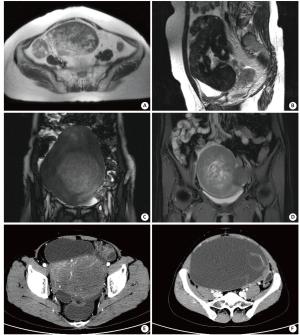
图 1 影像学表现
A: 右侧卵巢实性卵泡膜细胞瘤, 边界清, 可见外突结节, T2WI示肿瘤信号不均匀, 部分呈地图样高信号, 部分呈低信号; B: 左侧卵巢囊实性卵泡膜细胞瘤, 实性为主, 椭圆形, 可见脐凹征, 实性部分在T2WI上呈明显低信号, 内见多发片状、裂隙状囊变区, 呈高信号; C: 右侧卵巢实性卵泡膜细胞瘤, 中央组织较疏松, 含水成份多, 而周围纤维组织较致密, 在T2WI像呈周围低信号, 中央高信号, 形似“荷包蛋”; 盆腔少量积液; D: 右侧卵巢实性卵泡膜细胞瘤, 边界清, T2WI脂肪抑制像示“荷包蛋征”, 左侧见一外突结节, 呈低信号; 盆腔少量积液; E: 左侧卵巢实性卵泡膜细胞瘤, 类椭圆形, 边界清, CT增强扫描示瘤内较多纤细小血管, 但肿瘤仅呈轻中度强化; 盆腔少量积液; F: 左侧卵巢囊实性卵泡膜细胞瘤, 绝大部分呈囊性, 仅左侧见少许实性部分, 囊内可见分隔, CT增强扫描示分隔中可见纤细小血管, 实性部分呈轻度强化.
-
[1] 赵燕风, 戴景蕊, 王小艺, 等. 卵巢卵泡膜细胞瘤的CT表现[J]. 放射学实践, 2010, 25(7): 780-3. doi: 10.3969/j.issn.1000-0313.2010.07.022 [2] 侯 岩, 叶兆祥, 李绪斌, 等. 卵巢纤维瘤和纤维卵泡膜细胞瘤的CT表现[J]. 临床放射学杂志, 2013, 32(1): 84-7. [3] 陈本宝, 徐勇飞, 王善军, 等. 卵巢卵泡膜细胞瘤的CT表现与病理对照分析[J]. 医学影像学杂志, 2010, 20(12): 1864-7. doi: 10.3969/j.issn.1006-9011.2010.12.034 [4] 韦进军, 黎军强, 黄 欣, 等. 卵巢纤维卵泡膜细胞瘤的MRI诊断[J]. 广西医学, 2014, 26(12): 1815-7. [5] Troiano RT, Lazz AK, Scou TL, et al. Fibroma and fib rothecomaof the ovary: Mr imaging findings[J]. Radiology, 1997, 204(3): 795-8. doi: 10.1148/radiology.204.3.9280262 [6] 张 彦, 戚晓渊, 王立侠, 等. 血清CA125联合CT、MRI对卵巢卵泡膜细胞瘤的诊断价值[J]. 中国综合临床, 2015, 31(8): 759-62. doi: 10.3760/cma.j.issn.1008-6315.2015.08.027 [7] Li XC, Zhang WD, Zhu GB, et al. Imaging features and pathologic characteristics of ovarian thecoma[J]. J Comput Assist Tomogr, 2012, 36(1): 46-53. doi: 10.1097/RCT.0b013e31823f6186 [8] 杨 梅, 郭顺林, 雷军强, 等. 卵巢卵泡膜细胞瘤MRI表现与病理[J]. 中国医学影像学杂志, 2014, 22(4): 285-8. doi: 10.3969/j.issn.1005-5185.2014.04.012 [9] 张 静, 王培军, 袁小东, 等. 卵巢卵泡膜细胞瘤的MRI表现与病理对照研究[J]. 中华放射学杂志, 2007, 41(11): 1217-9. doi: 10.3760/j.issn:1005-1201.2007.11.018 [10] 石甜甜, 丁建国, 缪小芬, 等. 卵巢性索间质肿瘤的影像诊断[J]. 实用肿瘤杂志, 2011, 26(2): 161-4. [11] Shinagare AB, Meylaerts LJ, Laury AR. MRI features of ovarian fibroma and fibrothecoma with histopathologic correlation[J]. Am J Roentgenol, 2012, 198(3): W296-303. doi: 10.2214/AJR.11.7221 [12] Vijayaraghavan GR, Lwvine D. Case 109: meigs syndrome[J]. Radiology, 2007, 242(3): 940-4. doi: 10.1148/radiol.2423040775 [13] 段小玲, 陈自谦, 钟 群, 等. 卵巢卵泡膜细胞瘤的MRI表现[J]. 中国CT和MRI杂志, 2017, 15(2): 83-5. doi: 10.3969/j.issn.1672-5131.2017.02.026 [14] 周 玮, 何 剑, 沈 建, 等. 卵巢纤维卵泡膜细胞瘤的影像学表现[J]. 中国医学科学院学报, 2015, 37(4): 378-83. doi: 10.3881/j.issn.1000-503X.2015.04.002 [15] 程虹. 乳腺及女性生殖器官肿瘤病理学和遗传学[M]. 北京: 北京人民卫生出版社, 2006: 185-6. [16] 郑少燕, 曾向廷, 吴先衡, 等. 卵巢卵泡膜细胞瘤的MRI诊断及鉴别诊断[J]. 影像诊断与介入放射学, 2014, 16(2): 117-20. doi: 10.3969/j.issn.1005-8001.2014.02.005 [17] 汪文斌, 马周鹏, 王 春. 卵巢纤维瘤MRI诊断和鉴别诊断[J]. 中国临床医学影像杂志, 2012, 23(2): 131-4. doi: 10.3969/j.issn.1008-1062.2012.02.019 [18] 庄 严, 张国福, 田晓梅, 等. 纤维卵泡膜细胞瘤的MRI表现及误诊分析[J]. 上海医学影像, 2012, 21(1): 21-4. doi: 10.3969/j.issn.1008-617X.2012.01.007 [19] Thomassin-Naggara I, Darai E, Nassar-Slaba J, et al. Value of dynamicenhanced magnetic resonance imaging for distinguishing between ovarian fibroma and subserous uterine leiomyoma[J]. J Comput Assist Tomogr, 2007, 31(2): 236-47. doi: 10.1097/01.rct.0000237810.88251.9e [20] Nocito AL, Sarancone S, Bacchi C, et al. Ovarian thecoma: clinicopathological analysis of 50 cases[J]. Annal Diagn Pathol, 2008, 12(1): 12-6. doi: 10.1016/j.anndiagpath.2007.01.011 [21] 仪孝臣, 赵瑞峰, 迟 磊. 卵巢卵泡膜细胞瘤MRI表现[J]. 牡丹江医学院学报, 2012, 33(2): 44-6. doi: 10.3969/j.issn.1001-7550.2012.02.024 [22] Choi JI, Park SB, Han BH, et al. Imaging features of complex solid and multicystic ovarian lesions: proposed algorithm for differential diagnosis[J]. Clin Imaging, 2016, 40(1): 46-56. doi: 10.1016/j.clinimag.2015.06.008 -


 下载:
下载:


