


Prenatal ultrasonic diagnosis and analysis of right aortic arch
-
摘要:
目的探讨胎儿右位主动脉弓(RAA)的典型超声声像图表现的诊断价值,以提高其产前诊断的准确性。 方法回顾性分析2014~2018年于4家医院诊断并在产后明确的RAA病例的声像图特征及其产后随访情况。 结果78例胎儿时期诊断为RAA的病例中,有37例在产后通过超声心动图检查或CT检查明确诊断,其中RAA合并迷走左锁骨下动脉(RAA-ALSA)24例,RAA合并镜像分支(RAA-MI)10例;在三血管-气管切面中出现“U”型结构的胎儿约占78.4%,其中RAA伴迷走左锁骨下动脉、左后位动脉导管(RAA/ALSA/LPDA)24例(82.4%),DAA 3例(10.3%),RAA伴镜像分支、左后位动脉导管(RAA/MI/LPDA)2例(6.9%)。出现“U”型结构的病例均伴有完全性血管环形成,而“V”形结构在37例胎儿中的显现率为5.4%,所有胎儿均未发现血管环形成。另外,有6例(16.2%)胎儿既不出现“U”型结构,也不出现“V”形结构,均为右位主动脉弓合并左前位动脉导管,且无血管环形成。此外,RAA与心内结构畸形具有一定相关性。12例合并心内畸形,其中不形成血管环的胎儿合并心内畸形率约66.7%,且多合并复杂心内畸形。形成血管环的右位主动脉弓合并心内畸形率约13.8%,主要与简单先天性心脏病相关。 结论研究发现三血管-气管切面出现“V”形结构仅仅意味着主动脉弓横部和动脉导管指向胸部的同一侧,而出现“U”形结构则是血管环形成的标,血管环形成与否也与是否合并心内畸形关系密切,为右位主动脉弓的产前诊断和判断预后提供指导意见。 Abstract:ObjectiveTo investigate the diagnostic value of typical ultrasound sonographic features of the right aortic arch (RAA) in the fetus andto improve the accuracy of prenatal diagnosis. MethodsEchocardiographic characteristics of thefetal RAA cases diagnosed and confirmed in our hospital from 2014 to 2018 were retrospectively analyzed. ResultsIn 78 consecutive fetuses with a diagnosis of RAA, 37 patients were diagnosed after birth by echocardiography or CT, including 24 cases of RAA with aberrant left subclavian artery (RAA - ALSA) and 10 cases of RAA with mirror image branching pattern (RAA‐MI). Fetal " U-shaped”appearance was found in 29/37 patients (82%) and all had a complete vascular ring, included 24 patients (82.4%) had RAA with left posterior ductus arteriosus (LPDA) and aberrant left subclavian artery (ALSA) (RAA/LPDA/ALSA), 3 patients (10.3%) with double aortic arch (DAA), 2 patients (6.9%) RAA with LPDA and mirror image branching (RAA/LPDA/MI). Fetal " V-shaped” appearance was found in 8 patients (5.4%) and all had RAA with right DA (RDA) not forming vascular rings. In addition, 6/37 (16.2%) fetuses didn’t have a " U-shaped” or " V-shaped” structure, found with left anterior ductusarteriosus and mirror image branching branching andwithout forming vascular rings. RAA was related with intracardiac structural abnormalities. There were 12 patients with a congenitalheart defectin, which " RAA forming vascular rings”was related with simple congenital heart diseases in 4/29 (13.8%). " RAA not forming vascular rings” was related with complex congenital heart diseases in 8/12 (66.7%). Conclusion" V-shaped” appearance only means that the transverse section of the aortic arch and the arterial catheter run together on the same side of the thorax, while " U-shaped” appearance is the sign of a vascular ring. RAA forming vascular ring or not is closely related to congenital heart diseases and provides guidance for prenatal diagnosis and prognosis of the right aortic arch. -
Key words:
- fetal echocardiography /
- vascular ring /
- right aortic arch /
- congenital heart disease
-

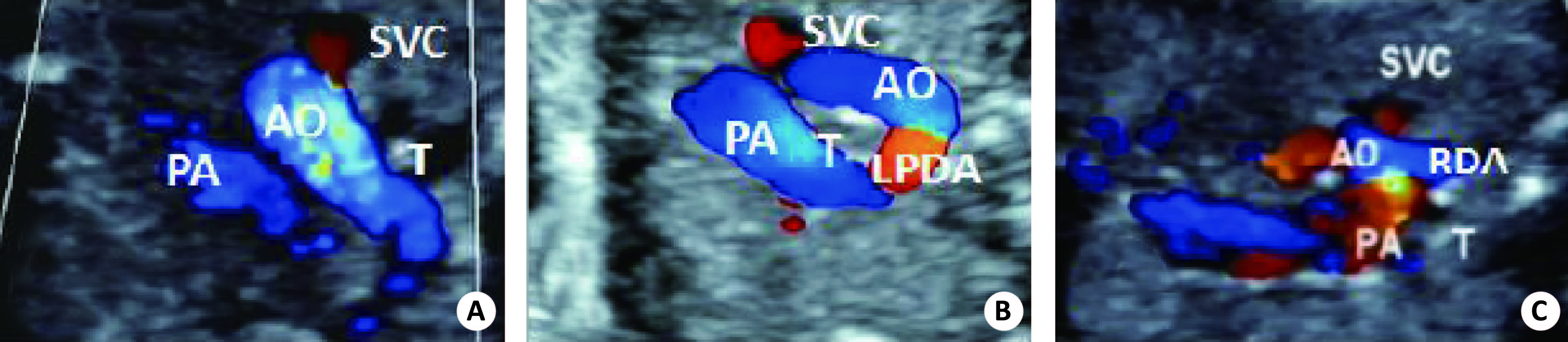
图 1 主动脉弓的典型超声图像
A:正常胎儿三血管-气管切面;B:右位主动脉弓合并左后位动脉导管的超声图像;C:右位主动脉弓合并右位动脉导管的超声图像;SVC:上腔静脉;AO:主动脉弓;T:气管;PA:肺动脉;LPDA:左后位动脉导管;RDA:右位动脉导管.
表 1 37例RAA胎儿的声像图表现、分支类型及是否形成血管环情况
分组 例数(%) 三血管-气管切面观 动脉导管位置 分支情况 例数(%) 迷走左锁骨下动脉 24(82.7) 右位主动脉弓形成完全血管环 29(78.4) “U”字型结构 左后动脉导管 双主动脉弓 3(10.3) 镜像分支 2(6.9) 右位主动脉弓不形成完全血管环 8(21.6) “V”字型结构 右位动脉导管 镜像分支 2(25.0) 不形成血管交汇结构 左前动脉导管 镜像分支 6(75.0)  下载: 导出CSV
表 2 右位主动脉弓病例特点
右位主动脉弓类型 例数 合并畸形种类 动脉导管位置 是否形成完全性血管环 RAA-ALSA 24 合并心内畸形 4 室间隔缺损4例 左后位动脉导管4例 是 不合并心内畸形 20 左后位动脉导管20例 是 DAA 3 合并心内畸形 0 不合并心内畸形 3 双主动脉弓3例 是 RAA-MI 10 合并心内畸形 8 法洛四联症4例 左前主动脉导管4例 否 右室双出口2例 右位动脉导管2例 否 肺动脉闭锁/室间隔缺损2例 左前动脉导管1例 否 左后位动脉导管1例 不合并心内畸形 2 左后位动脉导管1例 是 左前位动脉导管1例 否
下载: 导出CSV
-
[1] Edwards JE. Anomalies of the aortic arch system[J]. Birth Defects Orig Artic Ser, 1977, 13(3D): 47-63. [2] Achiron R, Rotstein Z, Heggesh J, et al. Anomalies of the fetal aortic arch: a novel sonographic approach to in-utero diagnosis[J]. Ultrasound Obstet Gynecol, 2002, 20(6): 553-7. doi: 10.1046/j.1469-0705.2002.00850.x [3] Peng R, Xie HN, Zheng J, et al. Fetal right aortic arch: associated anomalies, genetic anomalies with chromosomal microarray analysis, and postnatal outcome[J]. Prenat Diagn, 2017, 37(4): 329-35. doi: 10.1002/pd.v37.4 [4] 刘伟伟, 王岳恒, 王 菊. 产前超声诊断右位主动脉弓及其分支类型的价值[J]. 中华超声影像学杂志, 2018, 27(8): 674-7. doi: 10.3760/cma.j.issn.1004-4477.2018.08.008 [5] Yagel S, Arbel R, Anteby EY, et al. The three vessels and trachea view(3VT)in fetal cardiac scanning[J]. Ultrasound Obstet Gynecol, 2002, 20(4): 340-5. doi: 10.1046/j.1469-0705.2002.00801.x [6] Bravo C, Gamez F, Perez R, et al. Fetal aortic arch anomalies key sonographic views for their differential diagnosis and clinical implications using the cardiovascular system sonographic evaluation protocol[J]. J Ultrasound Med, 2016, 35(2): 237-42. doi: 10.7863/ultra.15.02063 [7] Yoo SJ, Min JY, Lee YH, et al. Fetal sonographic diagnosis of aortic arch anomalies[J]. Ultrasound Obstet Gynecol, 2003, 22(5): 535-46. doi: 10.1002/uog.897 [8] Gardiner H, Chaoui R. The fetal three-vessel and tracheal view revisited[J]. Semin Fetal Neonatal Med, 2013, 18(5): 261-8. doi: 10.1016/j.siny.2013.01.007 [9] 朱梦梦, 蔡爱露, 孙 悦, 等. 三血管气管切面在诊断胎儿右位主动脉弓中的应用价值分析[J]. 中国临床医学影像杂志, 2016, 27(7): 506-8, 511. [10] Lesieur E, Dabadie A, Pico H, et al. Segmental approach to congenital heart diseases: Principles and applications to prenatal imaging[J]. Gynecol Obstet Fertil, 2016, 44(7/8): 428-34. [11] 李雪蕾, 穆仲平, 胡克非, 等. 产前超声诊断胎儿右位主动脉弓18例分析[J]. 中国超声医学杂志, 2015, 31(6): 569-71. [12] 颜幸燕, 黄朝宁. 27例胎儿右位主动脉弓产前超声诊断分析[J]. 中国超声医学杂志, 2016, 32(4): 346-8. doi: 10.3969/j.issn.1002-0101.2016.04.019 [13] 郭艳霞, 肖 珍, 王丽敏, 等. 胎儿右位主动脉弓的产前超声诊断[J]. 广东医学, 2016, 37(11): 1675-7. [14] 马建芳, 邓学东, 潘 琦, 等. 右位主动脉弓胎儿超声心动图诊断分析[J]. 中华医学超声杂志:电子版, 2012, 18(10): 887-90. [15] 王新霞, 栗河舟, 王 铭, 等. 胎儿右位主动脉弓及合并畸形的超声诊断分析[J]. 中国妇幼保健, 2015, 35(6): 922-4. [16] Trobo D, Bravo C, Alvarez T, et al. Prenatal sonographic features of a double aortic arch literature review and perinatal management[J]. J Ultrasound Med, 2015, 34(11): 1921-7. doi: 10.7863/ultra.14.12076 [17] Miranda JO, Callaghan N, Miller O, et al. Right aortic arch diagnosed antenatally: associations and outcome in 98 fetuses[J]. Heart, 2014, 100(1): 54-9. doi: 10.1136/heartjnl-2013-304860 [18] Kobayashi M, Ando M, Wada N, et al. Outcomes following surgical repair of aortic arch obstructions with associated cardiac anomalies[J]. Eur J Cardiothorac Surg, 2009, 35(4): 565-8. doi: 10.1016/j.ejcts.2008.09.052 [19] Berg C, Bender F, Soukup M, et al. Right aortic arch detected in fetal Life[J]. Ultrasound Obstet Gynecol, 2006, 28(7): 882-9. doi: 10.1002/uog.3883 [20] 彭 软, 谢红宁, 周 祎, 等. 胎儿右位主动脉弓相关异常、遗传物质异常及预后[J]. 中国产前诊断杂志: 电子版, 2017, 19(2): 12-6. [21] Mcelhinney DB, Clark BJ, Weinberg PM, et al. Association of chromosome 22q11 deletion with isolated anomalies of aortic arch laterality and branching[J]. J Am Coll Cardiol, 2001, 37(8): 2114-9. doi: 10.1016/S0735-1097(01)01286-4 -

 点击查看大图
图(1) / 表(2)
计量
- 文章访问数: 1639
- HTML全文浏览量: 729
- PDF下载量: 7
- 被引次数: 0